

Unit 1: Basic Cell Functions
Cell structure, membrane transport, osmosis, cell cycle & apoptosis.
Prepared by the PharmaWallah student team. Verify results with your textbook or instructor. Report an error · Editorial policy
Unit 1: Basic Cell Functions
Learning Objectives
- Describe the physical structure of the cell, including the plasma membrane, cytoplasm, and major organelles.
- Distinguish between the composition and distribution of intracellular fluid and extracellular fluid and understand their significance.
- Explain the mechanisms of movement of molecules across cell membranes, including diffusion, active transport, co‑transport, and counter‑transport.
- Understand the basic principles of osmosis, osmotic pressure, osmotic equilibrium, and their physiological importance.
- Describe the processes of endocytosis, phagocytosis, pinocytosis, exocytosis, the cell cycle, and apoptosis.
Core Content
1. Overview of the Physical Structure of the Cell
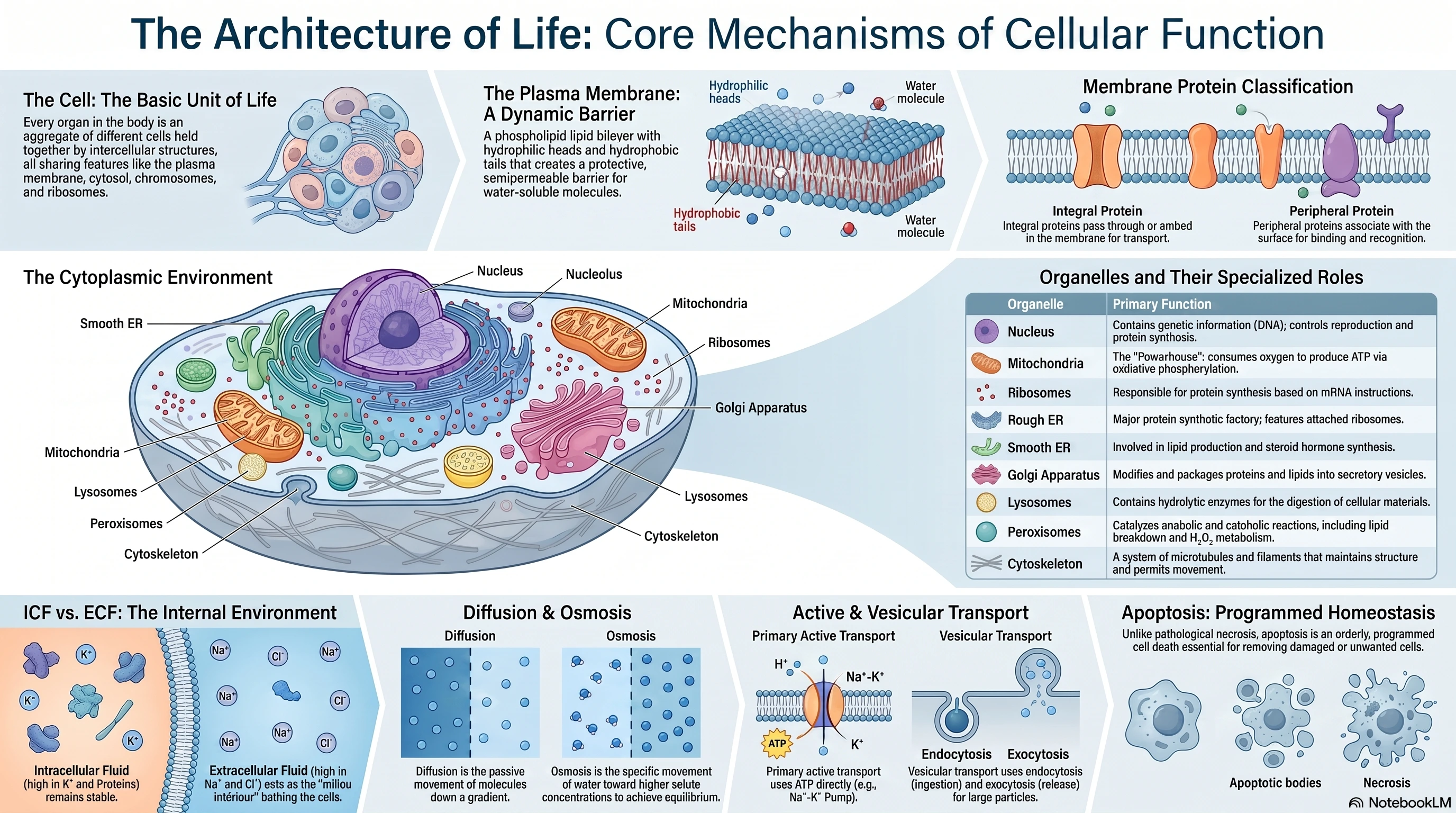
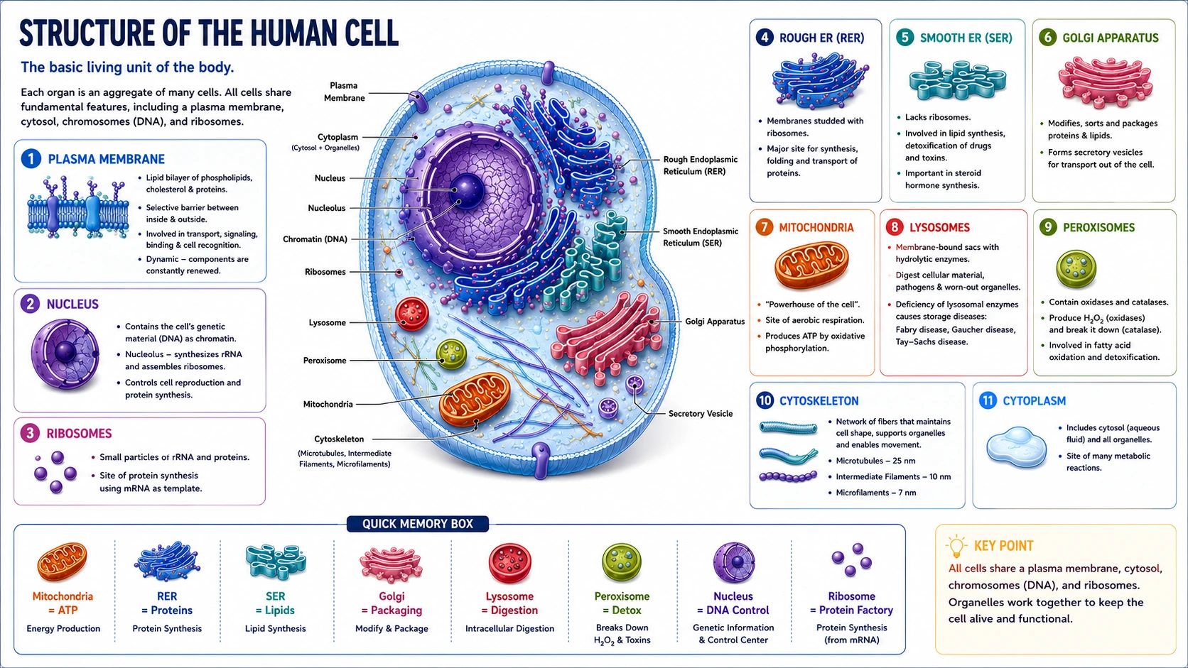
The cell is the basic living unit of the body. Each organ is an aggregate of many different cells held together by intercellular supporting structures. All cells share fundamental features, including a plasma membrane, cytosol, chromosomes, and ribosomes.
A. Cell Membrane (Plasma Membrane)
- Structure: The cell membrane is a lipid bilayer composed primarily of phospholipids with hydrophilic heads and hydrophobic tails, interspersed with proteins, glycolipids, and cholesterol. This arrangement forms a barrier that is generally impermeable to most water‑soluble molecules.
 The shape of the phospholipid molecule reflects its solubility properties: the hydrophilic "head" ends of the molecules are exposed to the aqueous environment, and the hydrophobic "tail" ends meet in the water‑poor interior of the membrane.
 - Functions: The membrane provides a barrier between intracellular compartments and the extracellular environment, protecting the cell and maintaining a stable internal environment. Within the membrane are proteins that function in cell transport, binding, and recognition. The membrane is a dynamic structure; its constituents are constantly renewed, and some proteins move laterally within it.
- Membrane Proteins: The membrane proteins are classified into two categories: integral proteins (intrinsic membrane proteins) which pass through or are embedded in one leaflet of the membrane, and peripheral proteins (extrinsic membrane proteins) which are associated with the inside or outside of the membrane. In epithelial cells, the enzymes in the cell membrane on the mucosal surface differ from those on the basal and lateral margins — the cells are polarized, making directional transport possible.
B. Cytoplasm and Organelles
The cell cytoplasm contains the cytosol (the aqueous component) and a variety of membrane‑bound organelles.
- Ribosomes: Responsible for protein synthesis based on instructions from mRNA.
- Endoplasmic Reticulum (ER):
- Smooth ER (SER): Involved in lipid production and steroid hormone synthesis.
- Rough ER (RER): Has attached ribosomes on its outer surface and is a major protein synthetic factory.
- Golgi Apparatus: Modifies lipids and proteins. Finished products are packaged as secretory vesicles for transport out of the cell.
- Lysosomes: Membrane‑bound organelles containing hydrolytic enzymes for digestion of cellular materials (e.g., ribonuclease, deoxyribonuclease, phosphatases, glycosidases, collagenase). When a lysosomal enzyme is congenitally absent, the lysosomes become engorged with the material the enzyme normally degrades, leading to lysosomal storage diseases such as Fabry disease, Gaucher disease, and Tay‑Sachs disease.
- Peroxisomes: Contain enzymes that can either produce H₂O₂ (oxidases) or break it down (catalases). They catalyze a variety of anabolic and catabolic reactions, including breakdown of lipids.
- Mitochondria: "Powerhouses" of the cell that consume oxygen in oxidative phosphorylation to produce ATP.
- Cytoskeleton: A system of fibers that maintains the structure of the cell and permits it to change shape and move. It is made up primarily of microtubules (25 nm), intermediate filaments (10 nm), and microfilaments (7 nm), along with proteins that anchor them.
- Nucleus: Contains the cell's genetic information (DNA). The DNA‑Genetic System controls cell reproduction and protein synthesis.
2. Extracellular Fluid and Intracellular Fluid
The body's water is distributed into two main fluid compartments: intracellular fluid (ICF) and extracellular fluid (ECF).
A. Intracellular Fluid (ICF)
- Definition: The fluid within cells, primarily the cytoplasm.
- Composition: High concentrations of potassium (K⁺), magnesium (Mg²⁺), phosphate (PO₄³⁻), and proteins; relatively lower concentrations of sodium (Na⁺), chloride (Cl⁻), and bicarbonate (HCO₃⁻). The ICF is relatively stable and does not readily adjust to rapid changes.
B. Extracellular Fluid (ECF)
- Definition: The fluid outside cells, which serves as the "internal environment" (milieu intérieur).
- Composition: High concentrations of sodium (Na⁺), chloride (Cl⁻), bicarbonate (HCO₃⁻), and proteins (in plasma); relatively lower concentrations of potassium (K⁺), magnesium (Mg²⁺), and phosphate.
- Subcategories: The ECF is further subcategorized into plasma (intravascular fluid), interstitial fluid (bathing the outside of cells), and transcellular fluid (e.g., cerebrospinal, gastrointestinal, intraocular fluids).
C. Significance
The composition and distribution of fluid spaces are maintained by active transport mechanisms, particularly the Na⁺‑K⁺ ATPase pump. The concentration of effective solutes determines the osmotic pressure gradient. Under normal conditions, the osmotic pressure of the ICF equates exactly with the plasma osmotic pressure.
3. Movement of Molecules Across Cell Membranes
Substances cross cell membranes by diffusion (passive), active transport (ATP‑requiring), and vesicular transport (endocytosis/exocytosis).
A. Diffusion
Diffusion is the passive movement of molecules down a concentration gradient, requiring no energy (ATP). Molecules can pass directly through the lipid bilayer if they are small and nonpolar, or via channels and carrier proteins (facilitated diffusion).
B. Active Transport
Active transport moves substances against their concentration gradient and requires energy, typically in the form of ATP.
- Primary Active Transport: The substance itself is moved using ATP directly. Example: The Na⁺‑K⁺ ATPase pump pumps three sodium ions out of a cell and two potassium ions in, cleaving one ATP in the process. This pump maintains cellular volume, maintains the membrane potential, and establishes chemical gradients used in secondary active transport.
C. Co‑Transport and Counter‑Transport
Secondary active transport couples the movement of one substrate down its concentration gradient to drive the movement of another substrate against its gradient.
- Co‑transport (Symport): Both substrates move in the same direction across the membrane. Example: Reabsorption of glucose in the kidney via the S‑GLUT transporter.
- Counter‑transport (Antiport): The two substrates move in opposite directions across the membrane. Example: The Na⁺/H⁺ exchanger in kidney cells.
4. Basic Principles of Osmosis, Osmotic Pressure, and Osmotic Equilibrium
Osmosis is the movement of water across a selectively permeable membrane from an area of lower solute concentration to an area of higher solute concentration. 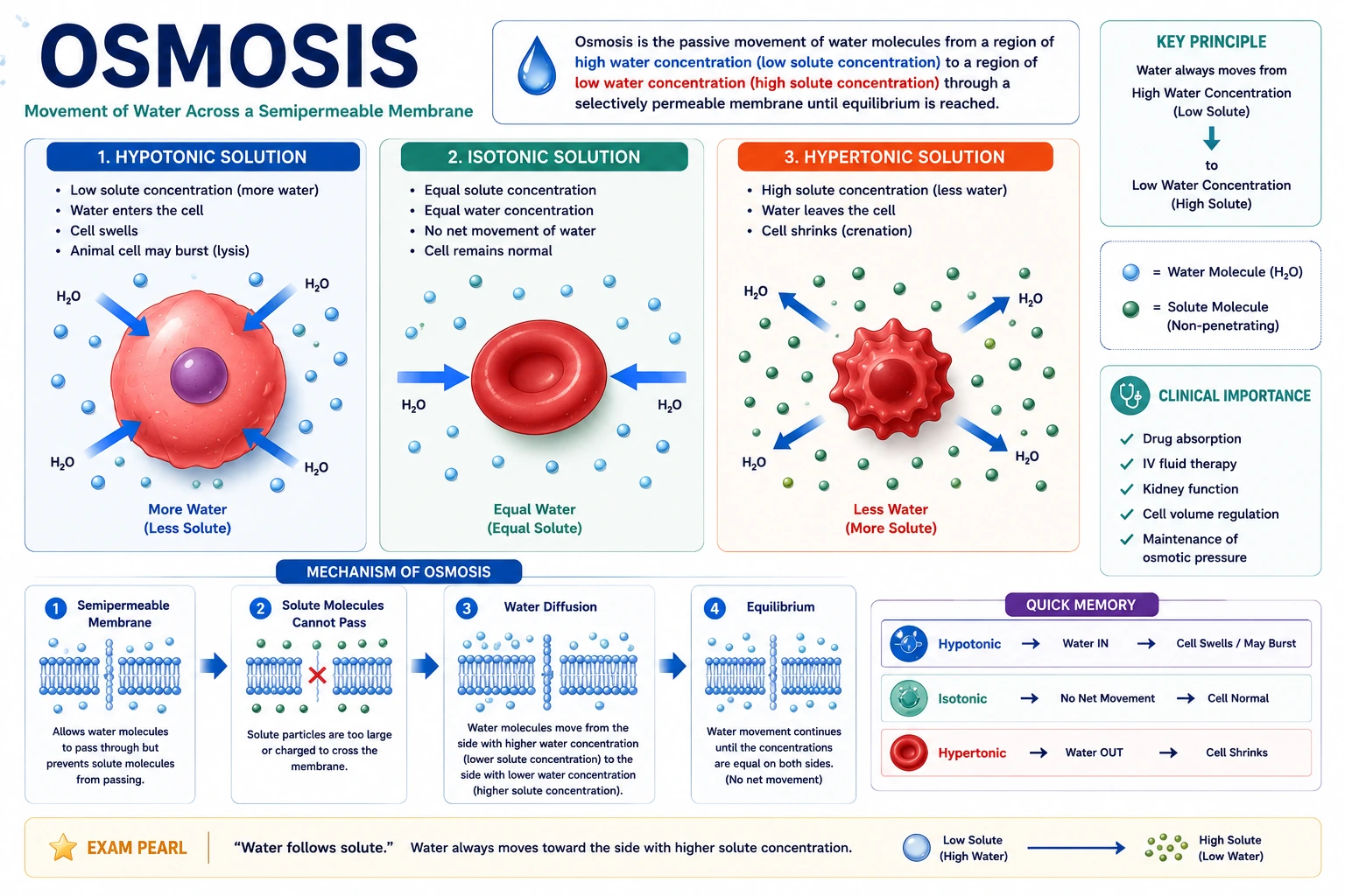 Water moves through the cell membrane by simple diffusion and is augmented by movement through water channels (aquaporins).
B. Osmotic Pressure and Equilibrium
- Osmotic Pressure: The pressure that must be applied to a solution to prevent the inward flow of water across a semipermeable membrane.
- Osmotic Equilibrium: The two fluid compartments of the body (ICF and ECF) are in osmotic equilibrium under normal conditions. The concentration of effective solutes (nonpermeable solutes like Na⁺ and Ca²⁺) determines the osmotic pressure gradient. Under steady‑state physiological conditions, the osmotic pressure of the ICF equates exactly with the plasma osmotic pressure.
- Significance: Cellular volume is dependent on the concentration gradient of effective solutes and water across the cell membrane. Cells shrink if the concentration of Na⁺ in the ECF is higher; cells swell if the concentration of Na⁺ in the ECF is lower.
5. Endocytosis and Exocytosis
A. Endocytosis
Endocytosis is the cellular ingestion of material from outside the cell, where the plasma membrane invaginates to form a vesicle.
- Pinocytosis: Non‑selective uptake of small particles within small vesicles ("cell drinking").
- Phagocytosis: Non‑selective uptake of large particles (>0.5 μm), such as microorganisms or dead cells ("cell eating").
- Receptor‑Mediated Endocytosis: Specific uptake of a substrate (ligand) through binding to specific receptors in membrane pits.
B. Exocytosis
Exocytosis is the process by which cells transport materials out of the cell. Vesicles fuse with the plasma membrane, releasing their contents into the extracellular fluid.
6. Cell Cycle and Apoptosis
A. Cell Cycle
The cell cycle is the series of events that leads to cell growth, DNA replication, and division into two daughter cells. The DNA‑Genetic System controls cell reproduction.
B. Apoptosis
Apoptosis is a form of programmed cell death that occurs in physiological conditions to maintain tissue homeostasis. It is essential for removing old, damaged, or unwanted cells in an efficient and orderly manner. This process is distinct from necrosis, which is pathological, uncontrolled cell death.
Tables
Table 1: Comparison of Intracellular and Extracellular Fluid
| Feature | Intracellular Fluid (ICF) | Extracellular Fluid (ECF) |
|---|---|---|
| Location | Within cells (cytoplasm) | Outside cells |
| High Concentration | K⁺, Mg²⁺, Phosphate, Proteins | Na⁺, Cl⁻, Bicarbonate, Proteins |
| Low Concentration | Na⁺, Cl⁻, Bicarbonate | K⁺, Mg²⁺, Phosphate |
| Maintenance | Na⁺‑K⁺ ATPase pump | Na⁺‑K⁺ ATPase pump |
Table 2: Comparison of Membrane Transport Mechanisms
| Mechanism | Energy Requirement | Direction | Description | Example |
|---|---|---|---|---|
| Simple Diffusion | Passive (none) | Down concentration gradient | Molecules pass through membrane/channels | O₂, CO₂, lipid‑soluble molecules |
| Facilitated Diffusion | Passive (none) | Down concentration gradient | Uses carrier proteins or channels | Glucose uptake via GLUT |
| Primary Active Transport | Active (ATP) | Against concentration gradient | Direct use of ATP to move substance | Na⁺‑K⁺ ATPase pump |
| Co‑transport (Symport) | Secondary | One substrate down gradient, one against | Both move same direction | Na⁺‑glucose cotransport in kidney |
| Counter‑transport (Antiport) | Secondary | One substrate down gradient, one against | Move opposite directions | Na⁺/H⁺ exchanger |
| Endocytosis (Phagocytosis/Pinocytosis) | Active (ATP) | Into cell (vesicle formation) | Cell engulfs particles/liquid | Macrophage engulfing bacteria |
| Exocytosis | Active (ATP) | Out of cell (vesicle fusion) | Vesicles fuse with membrane, release contents | Secretion of hormones, neurotransmitters |
Exam Angle
Short Answer Questions
- Define intracellular fluid and extracellular fluid. What are their key differences in composition?
- Differentiate between co‑transport and counter‑transport with examples.
- Explain the process of phagocytosis and its significance.
- What is apoptosis and how does it differ from necrosis?
- Describe the Na⁺‑K⁺ ATPase pump and its importance.
Essay Questions
- Discuss the mechanisms of transport across the cell membrane, including passive and active processes, and their physiological importance.
- Explain the basic principles of osmosis and osmotic equilibrium in the body. Describe the clinical significance of isotonic, hypotonic, and hypertonic solutions.
- Compare and contrast phagocytosis, pinocytosis, and receptor‑mediated endocytosis.
Viva / Short Notes
- Structure and function of the cell membrane.
- Lysosomal storage diseases.
- The cell cycle.
- Primary active transport vs. Secondary active transport.
- Cytoskeleton components.
Summary Box
- The cell is the basic unit of life, containing a plasma membrane (lipid bilayer), cytoplasm, and specialized organelles (nucleus, mitochondria, ER, Golgi, lysosomes) that perform distinct functions. The membrane is semipermeable and contains integral and peripheral proteins.
- Intracellular fluid (ICF) and extracellular fluid (ECF) are the two main body fluid compartments. ICF is high in K⁺ and phosphate, while ECF is high in Na⁺ and Cl⁻. The Na⁺‑K⁺ ATPase pump maintains these gradients.
- Movement across membranes occurs via diffusion (passive, down a concentration gradient) and active transport (ATP‑requiring, against a gradient). Primary active transport (e.g., Na⁺‑K⁺ ATPase) directly uses ATP; secondary active transport couples the movement of one molecule to another.
- Osmosis is the movement of water from low to high solute concentration. Osmotic equilibrium between ICF and ECF is essential for maintaining cell volume. Tonicity (isotonic, hypotonic, hypertonic) describes a solution's effect on cell volume and is vital in clinical fluid therapy.
- Endocytosis (phagocytosis and pinocytosis) brings substances into the cell via vesicle formation, while exocytosis releases substances from the cell. Apoptosis is programmed, non‑inflammatory cell death, distinct from necrosis.
References
- Hall, J. E., & Hall, M. E. (2021). Guyton and Hall Textbook of Medical Physiology (14th ed.). Elsevier.
- Barrett, K. E., Barman, S. M., Boitano, S., & Brooks, H. L. (2019). Ganong's Review of Medical Physiology (26th ed.). McGraw‑Hill Education.
- Boron, W. F., & Boulpaep, E. L. (2017). Medical Physiology (3rd ed.). Elsevier.
- Sherwood, L. (2016). Human Physiology: From Cells to Systems (9th ed.). Cengage Learning.
- Costanzo, L. S. (2018). Physiology (6th ed.). Elsevier.
End of unit 1 of 7
Check what you've learned
5 quick questions from this unit. They're optional: try them if you like, then mark the unit as read and move on.