

Unit 3: Circulation
Blood flow, haemodynamics, and regulation of circulation.
Prepared by the PharmaWallah student team. Verify results with your textbook or instructor. Report an error · Editorial policy
Unit 3: Circulation
Learning Objectives
- Describe the basic theory of circulatory function, including pressure, flow, and resistance relationships.
- Explain the structure and function of arteries, arterioles, capillaries, and veins.
- Define blood pressure, total peripheral vascular resistance, and pulmonary vascular resistance.
- Describe clinical methods of measuring systolic and diastolic blood pressures.
- Explain the arterial pressure baroreceptor reflex and the renin‑angiotensin system.
- Define hematocrit and describe the lymphatic system and formation of lymph.
Core Content
1. Basic Theory of Circulatory Function
The circulatory system is a closed system of tubes (vessels) that transports blood from the heart to tissues and back. Its fundamental function is to maintain adequate tissue perfusion, ensuring delivery of oxygen and nutrients and removal of metabolic waste. This function is governed by basic principles of hemodynamics analogous to electrical circuits.
A. Flow, Pressure, and Resistance Relationship
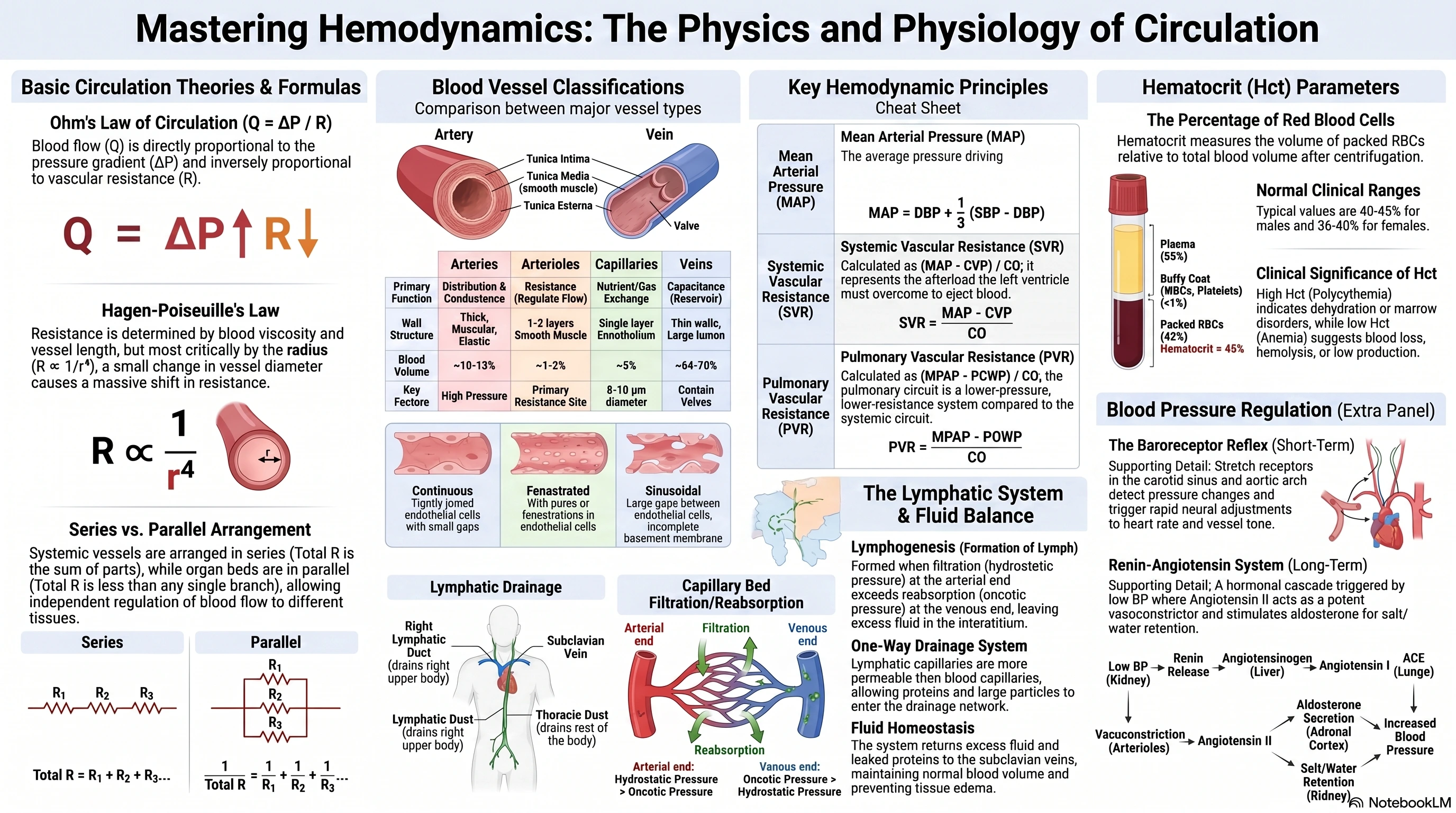
Ohm's Law Analogy: Blood flow (Q) through a vessel is determined by two factors: the pressure gradient (ΔP) and the resistance to flow (R).
Formula: Q = ΔP / R
Where:
- Q = Blood flow (cardiac output for the systemic circulation)
- ΔP = Pressure gradient (difference between upstream and downstream pressure, e.g., MAP – CVP for systemic circulation)
- R = Vascular resistance
Meaning: Blood flows from an area of higher pressure to an area of lower pressure. The greater the pressure difference, the greater the flow. The greater the resistance, the lower the flow. This is the most important concept in understanding vascular function.
B. Hagen‑Poiseuille's Law
This equation describes the factors that determine resistance to flow in a rigid tube.
Formula: R ∝ (η * L) / r⁴
Where:
- η = Viscosity of the blood
- L = Length of the vessel
- r = Radius of the vessel
Significance: The radius is the most powerful determinant of vascular resistance because it is raised to the fourth power. A small change in the diameter of an arteriole causes a large change in resistance and, consequently, blood flow. Vasoconstriction (decreased radius) increases resistance, while vasodilation (increased radius) decreases resistance.
C. Arrangement of Vasculature
- Series Arrangement: Vessels are arranged end‑to‑end (e.g., arteries → arterioles → capillaries → venules → veins). In a series, the flow is the same through all segments, but the pressures drop sequentially. Total resistance in series is the sum of individual resistances:
R_total = R₁ + R₂ + R₃ … - Parallel Arrangement: Vascular beds (e.g., to the kidneys, brain, muscles) are arranged in parallel. This arrangement allows for independent regulation of blood flow to different organs. In parallel, the total resistance is less than the resistance of any individual branch, allowing the system to handle high flow:
1/R_total = 1/R₁ + 1/R₂ + 1/R₃ …
2. Structure and Function of Blood Vessels
Blood vessels have three distinct layers, or tunics, with the relative thickness and composition varying by vessel type.
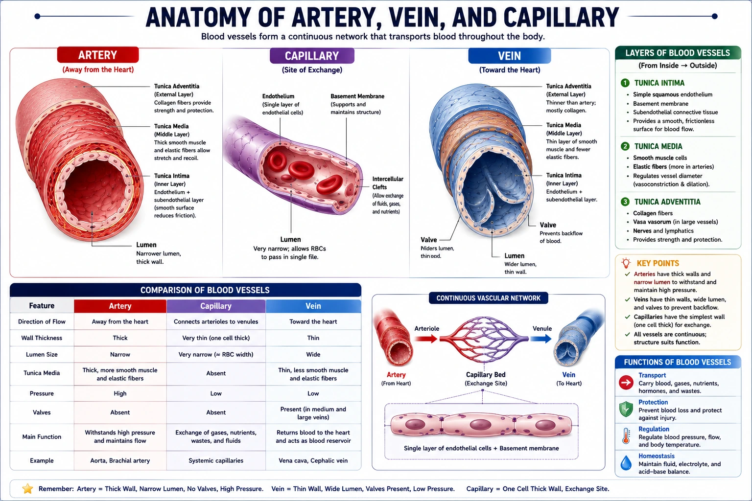
- Tunica Intima (Internal): The innermost layer, consisting of a simple squamous epithelium (endothelium) that lines the lumen. It provides a smooth, friction‑reducing surface.
- Tunica Media (Middle): The thickest layer in arteries, composed primarily of smooth muscle and elastic fibers. It regulates vessel diameter (vasoconstriction/vasodilation).
- Tunica Externa (Adventitia): The outermost connective tissue layer that anchors the vessel to surrounding tissues. It contains vasa vasorum (vessels that supply the vessel wall) and nervi vasorum (nerves).
A. Arteries
Carry blood away from the heart. They have thick, muscular, elastic walls designed to withstand and maintain high pressure.
- Elastic (Conducting) Arteries (e.g., aorta): The largest arteries, with abundant elastic fibers in the tunica media. They "conduct" blood and dampen pressure fluctuations through their elastic recoil.
- Muscular (Distributing) Arteries (e.g., femoral artery): Have more smooth muscle and distribute blood to specific organs.
- Arterioles: The smallest arteries, with only 1‑2 layers of smooth muscle in the tunica media. They are the primary resistance vessels. Their constriction or dilation controls blood flow into capillary beds and is the main determinant of total peripheral resistance.
B. Capillaries
The smallest blood vessels (8‑10 µm diameter) with walls composed of only a single layer of endothelial cells (tunica intima). They are the sites of exchange of nutrients, gases, and wastes between blood and tissues. Capillary distribution is related to metabolic activity of tissues.
Classification of Capillaries:
- Continuous: Most common; found in muscle and nervous tissue. Continuous basement membrane and tight junctions; transport requires carrier‑mediated transport.
- Fenestrated: Have pores (fenestrae) in the endothelial wall; found in endocrine glands, pancreas, and intestines.
- Sinusoidal (Discontinuous): Large fenestrae with incomplete basement membrane; found in liver, spleen, bone marrow. Allows for freer exchange of large molecules and cells.
C. Veins
Carry blood toward the heart. They are low‑pressure vessels with thinner walls, less smooth muscle, and larger lumens than arteries.
- Capacitance Vessels: Veins are distensible and hold approximately 70% of the total blood volume at any given time, serving as a blood reservoir.
- Venous Valves: Many medium and large veins (especially in the limbs) contain valves that prevent backflow of blood due to gravity. These are important for venous return.
3. Blood Pressure, Total Peripheral Vascular Resistance, and Pulmonary Vascular Resistance
A. Blood Pressure
Blood pressure is the force exerted by blood against the walls of the arteries. It is generated by the pumping of the heart and the resistance of the vessels. Normal values are around 120/80 mmHg.
- Systolic Blood Pressure (SBP): The peak arterial pressure during ventricular systole (contraction), reflecting the force of the heart's contraction.
- Diastolic Blood Pressure (DBP): The minimum arterial pressure during ventricular diastole (relaxation), reflecting the resistance of the peripheral vessels.
- Pulse Pressure (PP): The difference between systolic and diastolic pressure (
PP = SBP – DBP), reflecting the stroke volume and arterial compliance. - Mean Arterial Pressure (MAP): The average pressure in the arteries over the cardiac cycle. It is the main driving force for organ blood flow.
- Formula:
MAP = DBP + 1/3 (SBP – DBP)
- Formula:
B. Total Peripheral Vascular Resistance (TPR/SVR)
- Definition: The resistance to blood flow offered by all of the systemic vasculature. It is primarily determined by the degree of arteriolar constriction. Vasoconstriction increases SVR, and vasodilation decreases SVR.
- Calculation: SVR represents the afterload for the left ventricle.
SVR = (MAP – CVP) / CO(in Wood units or dyn·s·cm⁻⁵)- Where MAP = Mean Arterial Pressure; CVP = Central Venous Pressure; CO = Cardiac Output.
C. Total Pulmonary Vascular Resistance (PVR)
- Definition: The resistance to blood flow offered by the pulmonary vasculature. It is lower than SVR because the pulmonary circulation is a lower pressure, lower resistance system.
PVR = (MPAP – PCWP) / CO- Where MPAP = Mean Pulmonary Arterial Pressure; PCWP = Pulmonary Capillary Wedge Pressure; CO = Cardiac Output.
4. Clinical Methods of Measuring Systolic and Diastolic Blood Pressures
The most common non‑invasive method is auscultatory sphygmomanometry.
Procedure:
- An inflatable cuff is placed around the upper arm over the brachial artery.
- The cuff is inflated to a pressure above the expected systolic pressure, occluding the artery and stopping blood flow.
- A stethoscope is placed over the brachial artery, and the cuff is gradually deflated while listening for sounds (Korotkoff sounds).
- Systolic Pressure: The pressure at which the first clear, tapping Korotkoff sound appears, indicating blood is starting to flow through the partially occluded artery.
- Diastolic Pressure: The pressure at which the sounds disappear, indicating the artery is no longer occluded and blood flow is continuous.
Pharmacy Note: Accurate blood pressure measurement is essential for diagnosing hypertension and monitoring antihypertensive drug therapy (e.g., ACE inhibitors, ARBs, beta‑blockers, calcium channel blockers).
5. Arterial Pressure and Baroreceptor Reflex
Mean Arterial Pressure (MAP) is tightly regulated to ensure adequate tissue perfusion. The baroreceptor reflex is a rapid, short‑term neural mechanism (acting within seconds) to correct sudden changes in blood pressure.
Key Components:
- Baroreceptors: Stretch receptors located in the walls of the carotid sinus and the aortic arch. They respond to changes in arterial pressure. Increased pressure stretches them, increasing their firing rate.
- Afferent Pathways: Signals travel via the glossopharyngeal (CN IX, from carotid sinus) and vagus (CN X, from aortic arch) nerves to the nucleus tractus solitarius (NTS) in the medulla oblongata.
- Central Processing: The NTS integrates the signal and relays it to autonomic centers.
- Efferent Pathways:
- Parasympathetic (Cardioinhibitory) Center: Activated by increased pressure, it sends signals via the vagus nerve to the heart to decrease heart rate and contractility.
- Sympathetic (Vasomotor and Cardioacceleratory) Center: Inhibited by increased pressure, reducing sympathetic outflow to the heart and blood vessels.
Example: Response to Hypotension A drop in MAP reduces baroreceptor stretch and firing rate.
- This reduces the inhibition of the sympathetic center and decreases parasympathetic activity.
- Result:
- Sympathetic activation causes:
- Vasoconstriction → increases TPR, helping to raise MAP.
- Increased heart rate and contractility → increases Cardiac Output.
- Venoconstriction → increases venous return, supporting CO.
- Parasympathetic withdrawal further increases heart rate.
- Sympathetic activation causes:
These compensatory mechanisms work to restore MAP back to its normal set point.
Modulation: The renin‑angiotensin system (Angiotensin II) can modulate the baroreflex by resetting the reflex to a higher pressure and reducing its sensitivity.
6. Renin‑Angiotensin System (RAS)
The RAS is a long‑term (hours to days) regulator of blood pressure and blood volume. It is a major target for antihypertensive drugs.
Pathway:
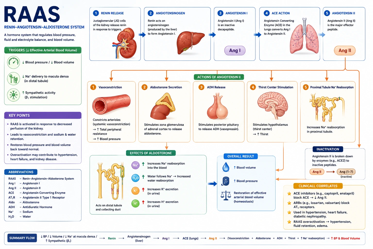
- Renin Release: Low blood pressure, decreased sodium delivery to the kidney, or sympathetic stimulation causes the kidneys to release the enzyme renin.
- Angiotensin I Formation: Renin acts on a plasma protein called angiotensinogen (produced by the liver), converting it to angiotensin I (inactive).
- Angiotensin II Formation: Angiotensin I is converted to Angiotensin II by Angiotensin‑Converting Enzyme (ACE), primarily in the lungs.
- Actions of Angiotensin II (a potent vasoconstrictor):
- Direct Vasoconstriction: Increases TPR.
- Stimulates Aldosterone Release: From the adrenal cortex, leading to sodium and water retention by the kidneys, increasing blood volume.
- Stimulates ADH (Vasopressin) Release: From the pituitary, increasing water reabsorption by the kidneys.
- Sympathetic Activation: Enhances sympathetic outflow.
- Central Effects: In the brain, it stimulates thirst and can reset the baroreflex to maintain a higher pressure.
Pharmacy Relevance: Drugs targeting this system include:
- ACE inhibitors (e.g., lisinopril, enalapril) – block conversion of Ang I to Ang II.
- Angiotensin Receptor Blockers (ARBs) (e.g., losartan, valsartan) – block AT₁ receptors.
- Renin inhibitors (e.g., aliskiren) – inhibit renin directly.
7. Hematocrit (Hct)
Definition: Hematocrit is the percentage of the total blood volume that is made up of red blood cells (erythrocytes).
Measurement: A blood sample is spun in a centrifuge; the heavier RBCs pack at the bottom, and the hematocrit is the ratio of the packed RBC volume to the total blood volume.
Clinical Significance:
- Normal Values: Approximately 40–45% for males and 36–40% for females.
- Increased Hct (Polycythemia): Can indicate dehydration (relative increase) or a bone marrow disorder.
- Decreased Hct (Anemia): Can indicate blood loss, low RBC production, or hemolysis.
8. Lymph Channels of the Body and Formation of Lymph
The lymphatic system is a low‑pressure, one‑way drainage system that parallels the venous system.
A. Lymphatic Vessels
- Lymphatic Capillaries: Blind‑ended, thin‑walled vessels that are found in most tissues. They are more permeable than blood capillaries and allow interstitial fluid, proteins, and large particles to enter.
- Lymphatic Vessels: Formed from the convergence of capillaries. They have valves to prevent backflow and are found in abundance in the skin, muscles, and digestive tract.
- Lymph Trunks and Ducts: These eventually merge into the right lymphatic duct (draining the right upper quadrant) and the thoracic duct (draining the rest of the body), which empty into the venous system (subclavian veins).
B. Formation of Lymph (Lymphogenesis)
Lymph is the fluid that enters the lymphatic capillaries from the interstitial space.
Mechanism: The formation of lymph is driven by a balance of forces at the capillary bed.
- Filtration: Hydrostatic pressure from the arterial end of the capillary forces fluid (water and small solutes) out of the blood and into the interstitial space.
- Reabsorption: Oncotic pressure (from plasma proteins) draws some of this fluid back into the venous end of the capillary.
- Excess Fluid: The remaining fluid, along with any leaked proteins, bacteria, or cell debris, is drained into the lymphatic capillaries.
- Function: The lymphatic system returns this fluid (now called lymph) to the bloodstream, maintaining normal blood volume and preventing edema.
Tables
Table 1: Comparison of Blood Vessel Types
| Feature | Arteries | Arterioles | Capillaries | Veins |
|---|---|---|---|---|
| Direction of Flow | Away from heart | Away from heart | Between arterioles/venules | Toward heart |
| Primary Function | Distribution, conductance | Resistance, regulate flow | Exchange of substances | Capacitance, return blood |
| Wall Thickness | Thickest | Thick (relatively) | Thin (single layer) | Thinner |
| Tunica Media | Most prominent (smooth muscle, elastic) | 1‑2 layers smooth muscle | Absent | Thin, less smooth muscle |
| Tunica Externa | Present, may have vasa vasorum | Present | Absent | Thickest layer in veins |
| Lumen | Small, round | Small | Very small (8‑10 µm) | Large, often flattened |
| Valves | Absent | Absent | Absent | Present in many (esp. limbs) |
| Blood Volume | ~10‑13% | ~1‑2% | ~5% | ~64‑70% |
Table 2: Key Hemodynamic Formulas
| Variable | Formula | Significance |
|---|---|---|
| Blood Flow (Q) | Q = ΔP / R | Flow is proportional to pressure gradient and inversely proportional to resistance. |
| Vascular Resistance (R) | R ∝ (η * L) / r⁴ | Small changes in vessel radius (r) cause large changes in resistance. |
| Systemic Vascular Resistance (SVR) | SVR = (MAP – CVP) / CO | The primary determinant of SVR is arteriolar constriction. |
| Pulmonary Vascular Resistance (PVR) | PVR = (MPAP – PCWP) / CO | A measure of resistance in the lower‑pressure pulmonary circuit. |
| Mean Arterial Pressure (MAP) | MAP = DBP + 1/3 (SBP – DBP) | The average pressure driving organ blood flow. |
Exam Angle
Short Answer Questions
- Define a baroreceptor and describe the baroreceptor reflex in response to a sudden drop in blood pressure.
- Explain the Hagen‑Poiseuille Law and its clinical significance.
- What is the function of venous valves?
- Differentiate between systemic vascular resistance and pulmonary vascular resistance.
- How is a hematocrit measured, and what clinical information does it provide?
Essay Questions
- Describe the structure and function of the three different types of blood vessels (arteries, capillaries, and veins), comparing their key anatomical features.
- Explain the short‑term neural and long‑term hormonal mechanisms that regulate blood pressure, focusing on the baroreceptor reflex and the renin‑angiotensin‑aldosterone system.
- Discuss the relationship between pressure, flow, and resistance in the cardiovascular system, explaining how total peripheral resistance and cardiac output influence mean arterial pressure.
Viva / Short Notes
- Korotkoff Sounds.
- The Lymphatic System and Lymph Formation.
- Factors affecting total peripheral resistance.
- Renin‑Angiotensin System as a drug target.
- Baroreceptor reflex pathway.
Summary Box
- The basic theory of circulation is governed by the relationship Flow = ΔP / R. The Hagen‑Poiseuille Law (R ∝ 1/r⁴) shows that the vessel radius is the most critical determinant of vascular resistance.
- Blood vessels have three tunics (intima, media, externa). Arteries are thick, pressure‑resistant conducting vessels. Arterioles are the main resistance vessels. Capillaries are thin‑walled exchange vessels. Veins are large, distensible capacitance vessels that hold most of the blood volume and contain valves.
- Blood Pressure is measured as systolic/diastolic pressure. Mean Arterial Pressure (MAP) is the perfusion pressure:
MAP = DBP + 1/3 × PP. - SVR is the resistance in the systemic circuit (
SVR = (MAP – CVP) / CO), primarily set by arterioles. PVR is the analogous resistance in the low‑pressure pulmonary circuit. - Blood pressure is regulated by the rapid baroreceptor reflex (neural, acting via the medulla to adjust HR and TPR) and the slower renin‑angiotensin system (hormonal, acting via Angiotensin II and aldosterone to control blood volume and resistance).
- Hematocrit is the percentage of blood volume composed of red blood cells, a key marker in assessing anemia or polycythemia.
- Lymph is formed from excess interstitial fluid driven by filtration and hydrostatic/oncotic forces at the capillary bed. It is returned to the blood via a system of lymphatic vessels (with lymph nodes), maintaining fluid balance.
References
- Hall, J. E., & Hall, M. E. (2021). Guyton and Hall Textbook of Medical Physiology (14th ed.). Elsevier.
- Barrett, K. E., Barman, S. M., Boitano, S., & Brooks, H. L. (2019). Ganong’s Review of Medical Physiology (26th ed.). McGraw‑Hill Education.
- Boron, W. F., & Boulpaep, E. L. (2017). Medical Physiology (3rd ed.). Elsevier.
- Sherwood, L. (2016). Human Physiology: From Cells to Systems (9th ed.). Cengage Learning.
- Katzung, B. G. (2018). Basic and Clinical Pharmacology (14th ed.). McGraw‑Hill Education.
End of unit 3 of 7
Check what you've learned
5 quick questions from this unit. They're optional: try them if you like, then mark the unit as read and move on.