

Unit 6: Digestion and Absorption of Food
Gastrointestinal physiology, enzymes & nutrient uptake.
Prepared by the PharmaWallah student team. Verify results with your textbook or instructor. Report an error · Editorial policy
Unit 6: Digestion and Absorption of Food
Learning Objectives
- Describe the composition, function, and regulation of gastrointestinal secretions (salivary, gastric, pancreatic, and biliary).
- Explain the nervous and hormonal regulation of gastric secretions.
- Describe the processes of mastication, deglutition, gastric emptying, and intestinal movements (peristalsis, segmentation, and mixing).
- Explain the enterohepatic circulation of bile and the process of defecation.
- Understand the overview of the enteric nervous system and its role in gastrointestinal function.
Introduction to the Digestive System
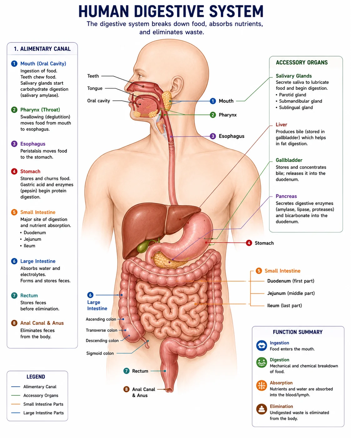
The digestive system breaks down food into absorbable nutrients, absorbs them into the bloodstream or lymph, and eliminates indigestible residues. It includes the gastrointestinal tract (GIT) and accessory organs (salivary glands, pancreas, liver, gallbladder).
Functions:
| Function | Description |
|---|---|
| Ingestion | Intake of food into the mouth |
| Propulsion | Movement of food through the tract (swallowing, peristalsis) |
| Mechanical digestion | Physical breakdown (chewing, churning, segmentation) |
| Chemical digestion | Enzymatic breakdown of macromolecules into absorbable units |
| Absorption | Passage of digested nutrients into blood or lymph |
| Defecation | Elimination of indigestible residues |
Layers of the Gastrointestinal Tract Wall:
| Layer | Composition | Function |
|---|---|---|
| Mucosa | Epithelium, lamina propria, muscularis mucosae | Secretion, absorption, protection |
| Submucosa | Connective tissue with blood vessels, lymphatics, submucosal (Meissner’s) plexus | Support, blood supply, secretion control |
| Muscularis externa | Inner circular and outer longitudinal smooth muscle; myenteric (Auerbach’s) plexus between them | Motility (peristalsis, segmentation) |
| Serosa / Adventitia | Connective tissue covered by mesothelium (visceral peritoneum) | Protection, reduces friction |
1. Gastrointestinal Secretions
A. Salivary Secretions
- Glands: Parotid (serous), submandibular (mixed), sublingual (mucous), and minor salivary glands.
- Volume: 0.8–1.5 L/day.
- Composition:
- Water (99.5%), electrolytes (Na⁺, K⁺, Cl⁻, HCO₃⁻)
- α‑Amylase (ptyalin): begins starch digestion (cleaves α‑1,4 bonds)
- Lingual lipase: minor fat digestion, active in stomach
- Mucus (mucin): lubrication, protection
- Lysozyme: antibacterial
- Lactoferrin: binds iron, inhibits bacteria
- IgA: immune protection
- Functions: Moistens food, begins carbohydrate/lipid digestion, cleanses mouth, buffers acids.
- Regulation:
- Parasympathetic (CN VII, IX): produces copious, watery, enzyme‑rich saliva (ACh via muscarinic receptors).
- Sympathetic: produces small volume of thick, mucinous saliva.
- Conditioned (sight, smell) and unconditioned (food in mouth) reflexes.
B. Gastric Secretions
- Glands: Gastric glands in fundus/body; pyloric glands in antrum.
- Volume: 1.5–2.5 L/day.
Cell Types and Their Secretions:
| Cell Type | Location | Secretion | Function |
|---|---|---|---|
| Parietal (oxyntic) cells | Body | HCl, Intrinsic Factor | Activates pepsinogen; kills bacteria; denatures proteins; IF binds B₁₂ |
| Chief (peptic) cells | Body | Pepsinogen (inactive) | Converted to pepsin by HCl; digests proteins |
| Mucous cells (surface, neck) | Throughout | Mucus, HCO₃⁻ | Protects mucosa; lubrication |
| G cells | Antrum | Gastrin | Stimulates acid secretion (hormonal) |
| D cells | Body, antrum | Somatostatin | Inhibits gastrin and acid secretion |
| ECL cells | Body | Histamine | Paracrine; stimulates acid secretion (via H₂ receptors) |
Mechanism of HCl Secretion (Parietal Cell):
- CO₂ + H₂O → H₂CO₃ (carbonic anhydrase).
- H⁺ pumped into lumen by H⁺/K⁺ ATPase (proton pump); K⁺ recycled.
- HCO₃⁻ exchanged for Cl⁻ at basolateral membrane (Cl⁻–HCO₃⁻ exchanger); Cl⁻ diffuses into lumen via channels.
- Net result: HCl secreted; HCO₃⁻ enters blood → alkaline tide after meal.
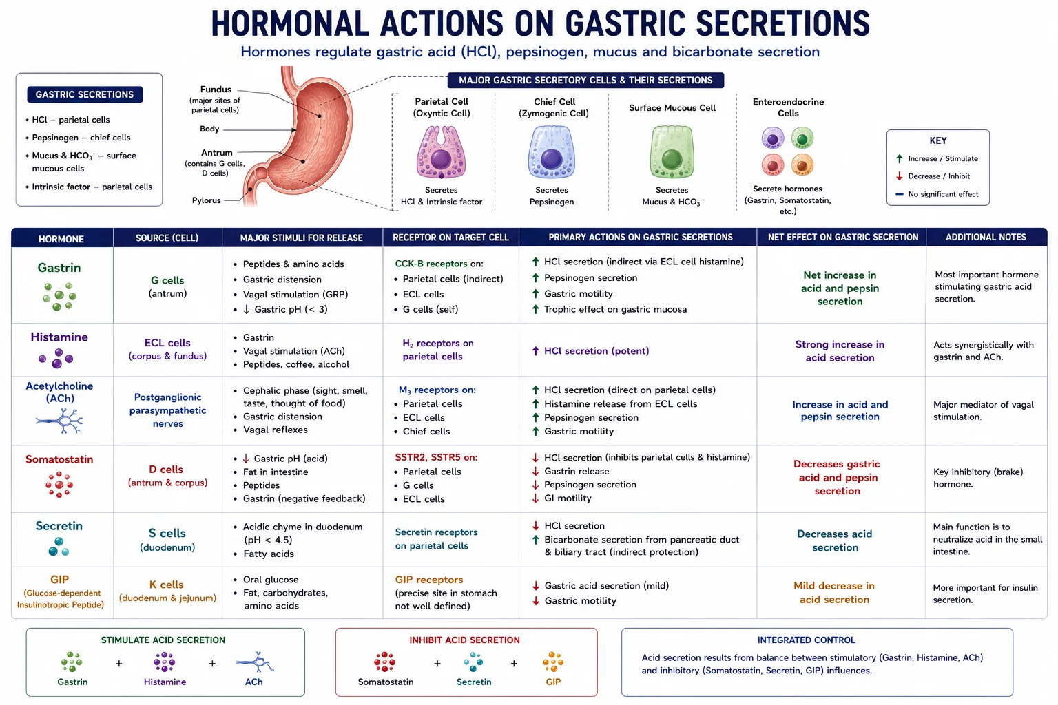
Phases of Gastric Secretion:
| Phase | % of Secretion | Stimulus | Mechanism |
|---|---|---|---|
| Cephalic | 30% | Sight, smell, taste, thought, chewing | Vagal stimulation → ACh → parietal cells + G cells (gastrin) |
| Gastric | 60% | Distension, peptides, amino acids | Vagovagal and local reflexes; gastrin release; histamine from ECL cells |
| Intestinal | 10% | Chyme in duodenum (initial excitation, then inhibition) | Amino acids/peptides → minor gastrin; fat, acid, hypertonicity → enterogastric reflex (secretin, CCK, GIP inhibit secretion and emptying) |
Inhibition: Acid (pH <3) → somatostatin from D cells → inhibits gastrin and parietal cells. Duodenal factors (fat, acid, hypertonicity) trigger the enterogastric reflex, releasing enterogastrones (secretin, CCK, GIP) that inhibit gastric secretion and motility.
C. Pancreatic Secretions
- Source: Exocrine pancreas (acinar cells – enzymes; duct cells – bicarbonate).
- Volume: 1.0–2.0 L/day.
Pancreatic Enzymes (secreted as zymogens where noted):
| Enzyme (Zymogen) | Activator | Substrate | Products |
|---|---|---|---|
| Trypsin (trypsinogen) | Enterokinase (duodenal brush border) | Proteins, peptides | Smaller peptides |
| Chymotrypsin (chymotrypsinogen) | Trypsin | Proteins, peptides | Smaller peptides |
| Elastase (proelastase) | Trypsin | Elastin | Peptides |
| Carboxypeptidase A/B (procarboxypeptidase) | Trypsin | Peptides (C‑terminal) | Amino acids |
| Pancreatic lipase | – | Triglycerides | Fatty acids + monoglycerides |
| Phospholipase A₂ (prophospholipase A₂) | Trypsin | Phospholipids | Fatty acid + lysophospholipid |
| Cholesterol esterase | – | Cholesterol esters | Cholesterol + fatty acid |
| α‑Amylase | – | Starch, glycogen | Maltose, maltotriose, limit dextrins |
| Ribonuclease, Deoxyribonuclease | – | RNA, DNA | Nucleotides |
-
Protection: Enzymes are stored as inactive precursors; trypsinogen activated only in duodenum by enterokinase; trypsin then activates other zymogens. Pancreatic trypsin inhibitor prevents premature activation.
-
Bicarbonate (from duct cells): neutralises gastric acid; provides optimal pH (7.5–8.0) for enzyme action.
Regulation:
- Secretin (from duodenal S cells, stimulated by acid): increases bicarbonate‑rich, enzyme‑poor secretion.
- CCK (from duodenal I cells, stimulated by fats & proteins): increases enzyme‑rich secretion.
- Vagal stimulation (cephalic & gastric phases): modest enzyme secretion.
D. Biliary Secretions and Enterohepatic Circulation
- Source: Liver hepatocytes; stored and concentrated (up to 10‑fold) in gallbladder.
- Volume: 0.6–1.2 L/day.
Composition:
- Bile salts (bile acids conjugated with glycine/taurine): cholic acid, chenodeoxycholic acid; secondary bile acids from bacteria. Function: emulsify fats, form micelles for lipid absorption.
- Bilirubin (conjugated): waste product excreted in faeces.
- Cholesterol: excreted; can form gallstones if supersaturated.
- Phospholipids (lecithin): solubilise cholesterol.
- Bicarbonate, electrolytes, water.
Enterohepatic Circulation:
- Bile salts are synthesised in liver from cholesterol and secreted into bile.
- Stored/concentrated in gallbladder; released into duodenum in response to CCK.
- ~95% of bile salts are actively reabsorbed in the terminal ileum.
- Returned via portal blood to liver, taken up, and re‑secreted (recycled 6–10 times/day).
- ~5% lost in faeces, replaced by new synthesis. Disruption (e.g., ileal resection, cholestyramine) causes bile salt deficiency, fat malabsorption, steatorrhoea, and fat‑soluble vitamin deficiencies (A, D, E, K).
Regulation:
- CCK: gallbladder contraction + sphincter of Oddi relaxation.
- Secretin: stimulates bicarbonate‑rich bile flow.
- Vagal stimulation: mild gallbladder contraction.
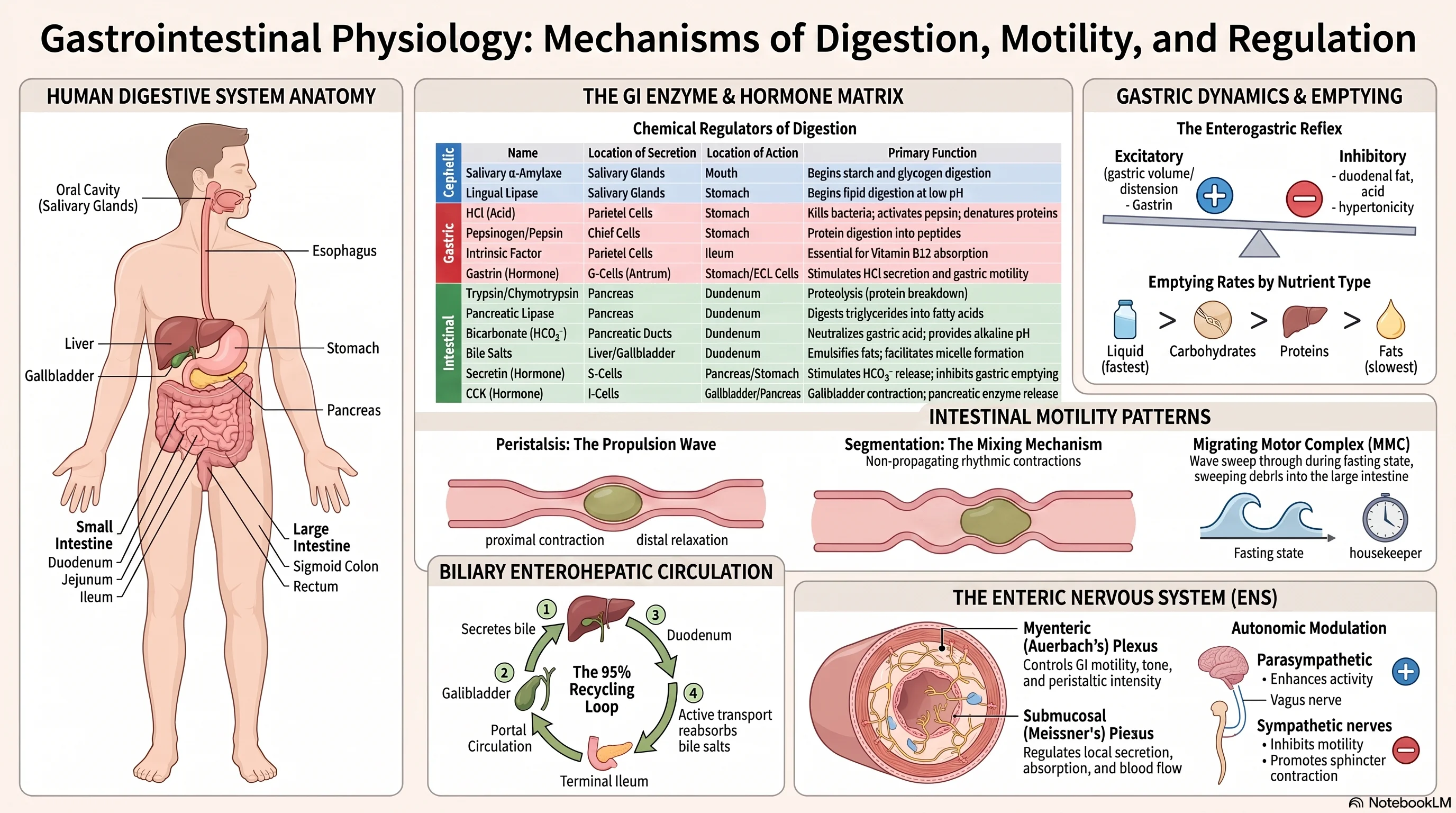
2. Gastrointestinal Hormones Overview
| Hormone | Source | Stimulus | Major Actions |
|---|---|---|---|
| Gastrin | G cells (antrum) | Peptides, amino acids, distension, ACh | ↑ gastric acid, motility, mucosal growth |
| CCK | I cells (duodenum) | Fatty acids, amino acids | ↑ pancreatic enzyme secretion, gallbladder contraction; ↓ gastric emptying |
| Secretin | S cells (duodenum) | Duodenal acid | ↑ pancreatic HCO₃⁻, ↓ gastric acid & emptying |
| GIP | K cells (duodenum) | Glucose, fats | ↑ insulin release (incretin effect); ↓ gastric acid |
| Motilin | M cells (duodenum) | Fasting | Initiates migrating motor complex (MMC) |
| Somatostatin | D cells (stomach, intestine, pancreas) | Acid | Paracrine inhibitor of gastrin, acid, pancreatic secretion, motility |
| Ghrelin | Stomach (fundus) | Fasting | ↑ appetite, gastric motility |
3. Motility: Mastication, Deglutition, and Gastric Emptying
A. Mastication (Chewing)
- Mechanical breakdown of food, increases surface area for enzymes, mixes with saliva.
- Controlled by brainstem (trigeminal motor nucleus); voluntary initiation, then rhythmic reflex.
B. Deglutition (Swallowing)
| Phase | Description | Control | Duration |
|---|---|---|---|
| Oral (voluntary) | Tongue pushes bolus into oropharynx | Cerebral cortex | ~1 s |
| Pharyngeal (involuntary) | Soft palate elevates, epiglottis covers airway, pharyngeal constrictors propel bolus; UES relaxes | Swallowing centre in medulla (CN V, IX, X, XII) | <1 s |
| Esophageal (involuntary) | Primary peristalsis (continuation); secondary peristalsis if needed; LES relaxes | Vagus (CN X); enteric NS | 5–10 s |
- LES: tonically contracted to prevent reflux; relaxes upon swallowing (VIP, NO).
- Disorders: Achalasia (failure of LES relaxation), GERD (incompetent LES).
C. Gastric Emptying
- Factors promoting emptying: gastric distension (stretch reflexes, gastrin), gastrin enhances motility.
- Factors inhibiting emptying (enterogastric reflex): duodenal acid (secretin), fat (CCK), hypertonicity, distension → inhibit gastric motility and close pylorus.
- Liquids empty faster than solids; carbohydrates > proteins > fats.
4. Intestinal Movements
| Movement | Description | Location | Function |
|---|---|---|---|
| Segmentation | Stationary, rhythmic contractions mixing contents | Small intestine (8–12/min) | Mixing; exposure to mucosa for absorption |
| Peristalsis | Propagating wave of contraction preceded by relaxation | Entire GI tract | Propulsion aborally |
| Migrating Motor Complex (MMC) | Strong peristaltic waves during fasting, repeating every 90–120 min | Stomach, small intestine | “Housekeeper” – clears undigested debris |
| Haustrations | Slow segmental contractions | Colon | Mixing; water absorption |
| Mass movements | Powerful, long peristaltic contractions, 3–4 times/day (often after meals – gastrocolic reflex) | Colon | Propel faeces toward rectum |
- Ileocecal sphincter: tonically contracted; relaxes with peristaltic wave; gastrin, CCK increase tone.
- Gastrocolic reflex: gastric distension → increased colonic motility (gastrin, CCK, neural).
5. Defecation
- Rectal distension by faeces → stretch receptors → parasympathetic (pelvic splanchnic, S2‑4) → contraction of sigmoid colon and rectum, relaxation of internal anal sphincter (involuntary).
- Voluntary control: external anal sphincter (skeletal muscle, pudendal nerve) can be consciously relaxed or contracted; Valsalva manoeuvre (increased intra‑abdominal pressure) assists.
- Reflex centres: spinal (sacral) and higher centres can facilitate or inhibit.
6. Overview of the Enteric Nervous System (ENS)
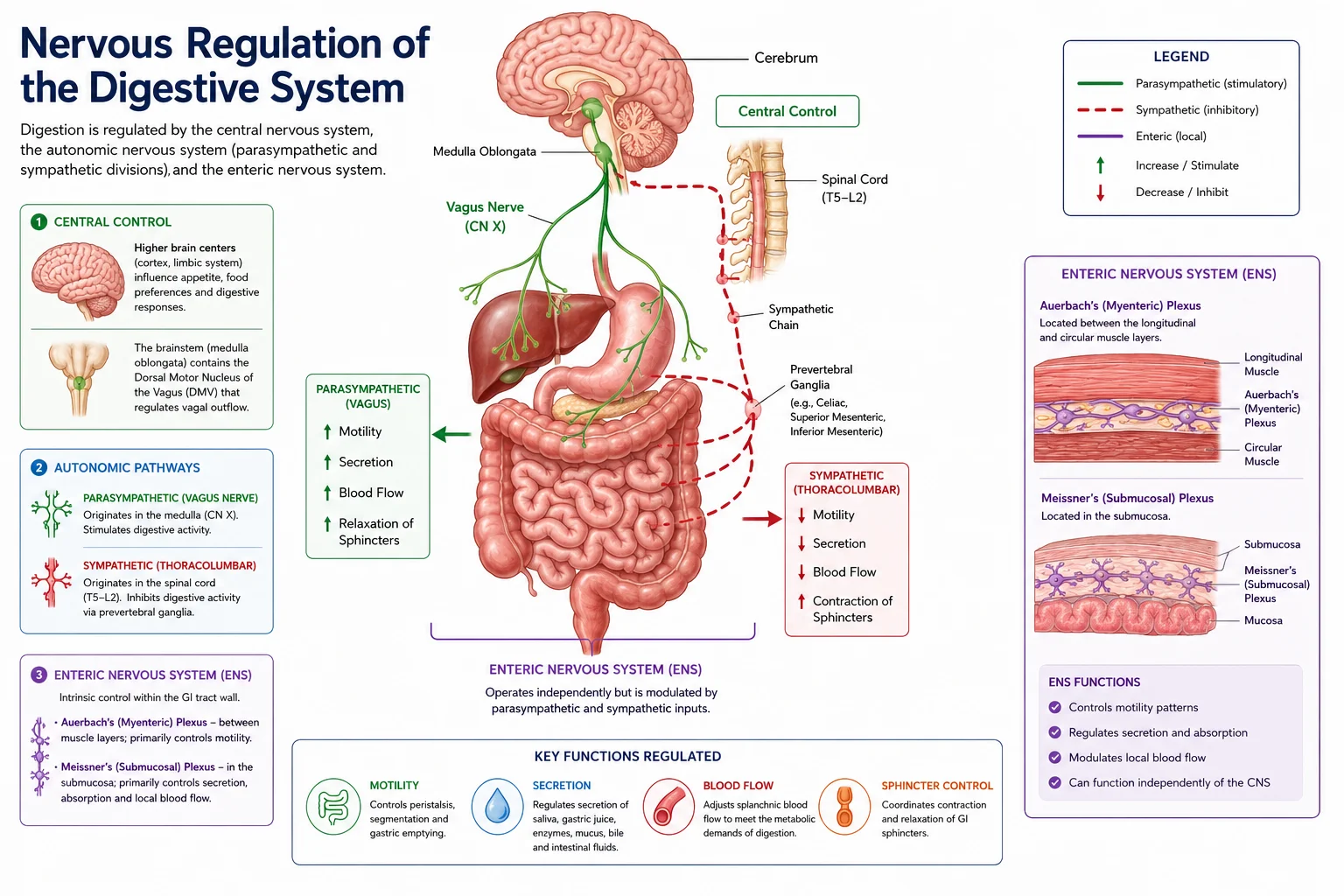
Often called the “second brain,” the ENS contains ~100 million neurons and can operate independently of the CNS, though modulated by autonomic input.
Plexuses:
- Myenteric (Auerbach’s) plexus: between longitudinal and circular muscle layers → controls motility (peristalsis, segmentation).
- Submucosal (Meissner’s) plexus: in submucosa → controls secretion, absorption, and blood flow.
Key Neurotransmitters:
| Neurotransmitter | Effect |
|---|---|
| Acetylcholine (ACh) | Excitatory (↑ motility, secretion) |
| Substance P | Excitatory |
| Nitric oxide (NO) | Inhibitory (smooth muscle relaxation) |
| Vasoactive intestinal peptide (VIP) | Inhibitory; stimulates secretion |
| Norepinephrine (sympathetic) | Inhibitory (relaxation via α₂ on cholinergic neurons) |
| Serotonin (5‑HT) | Modulates peristalsis; sensory transmission |
| Somatostatin | Inhibitory |
| Enkephalins | Inhibitory |
Functions: coordinates peristaltic reflex (ascending contraction, descending relaxation), regulates secretion and local blood flow, integrates sensory information.
Peristaltic Reflex: distension → sensory neurons → interneurons → excitatory motor neurons proximal (contraction) and inhibitory motor neurons distal (relaxation).
Clinical Relevance:
- Achalasia: loss of inhibitory neurons in esophagus → failure of LES relaxation.
- Hirschsprung’s disease: congenital absence of enteric ganglia in distal colon → functional obstruction.
- Diabetic gastroparesis: autonomic neuropathy affecting ENS.
- IBS: altered ENS function, visceral hypersensitivity.
Tables
Table 1: Comparison of Digestive Secretions
| Secretion | Daily Volume | pH | Key Components | Functions |
|---|---|---|---|---|
| Saliva | 0.8–1.5 L | 6.0–7.4 | α‑amylase, mucus, HCO₃⁻, lysozyme, IgA | Lubrication, starch/lipid digestion, antibacterial |
| Gastric juice | 1.5–2.5 L | 1.5–3.5 | HCl, pepsinogen, intrinsic factor, mucus | Protein digestion, kills bacteria, B₁₂ absorption |
| Pancreatic juice | 1.0–2.0 L | 7.5–8.0 | HCO₃⁻, proteases, lipase, amylase, nucleases | Neutralises acid, digests all macronutrients |
| Bile | 0.6–1.2 L | 7.0–8.0 | Bile salts, bilirubin, cholesterol, lecithin | Fat emulsification, excretion |
Table 2: Phases of Gastric Secretion
| Phase | % | Stimulus | Mechanisms |
|---|---|---|---|
| Cephalic | 30% | Sight, smell, taste, chewing | Vagal ACh → parietal cells + gastrin |
| Gastric | 60% | Distension, peptides | Vagovagal reflexes, gastrin, histamine |
| Intestinal | 10% | Duodenal chyme (initially stimulatory, then inhibitory) | Minor gastrin; enterogastric reflex (secretin, CCK, GIP) |
Table 3: Intestinal Motility Patterns
| Pattern | Location | Frequency | Function | Control |
|---|---|---|---|---|
| Segmentation | Small intestine | 8–12/min | Mixing, absorption | Myenteric plexus; interstitial cells of Cajal |
| Peristalsis | Entire GI tract | Variable | Propulsion | Enteric reflex |
| MMC | Stomach, small intestine | Every 90–120 min (fasting) | Clear debris | Motilin |
| Mass movements | Large intestine | 3–4/day | Propulsion to rectum | Gastrocolic reflex (gastrin, CCK, neural) |
Exam Angle
Short Answer Questions
- What are the components and functions of pancreatic juice?
- Describe the enterohepatic circulation of bile salts.
- Differentiate between peristalsis and segmentation.
- Summarise the three phases of gastric secretion.
- What is the function of the enteric nervous system?
Essay Questions
- Discuss the nervous and hormonal regulation of gastric secretion in all three phases.
- Explain the regulation of pancreatic secretion, including the roles of secretin and CCK.
- Describe the digestion and absorption of fats, highlighting the roles of bile salts, pancreatic lipase, and micelle formation.
- Outline the control of gastrointestinal motility, including gastric emptying, intestinal movements, and defecation.
Viva / Short Notes
- Enterohepatic circulation
- Defecation reflex
- Functions of bile salts
- Migrating motor complex (MMC)
- Composition and functions of gastric juice
- Hormonal regulation of pancreatic secretion
Summary Box
- GI secretions (saliva, gastric juice, pancreatic juice, bile) contain enzymes, electrolytes, and mucus for digestion and protection. Saliva begins carbohydrate/lipid digestion; gastric juice digests proteins and kills bacteria; pancreatic juice digests all macronutrients; bile emulsifies fats.
- Gastric secretion is controlled in three overlapping phases: cephalic (vagal), gastric (distension, peptides, gastrin), and intestinal (enterogastric reflex). Somatostatin provides negative feedback when pH is low.
- Gastric emptying is promoted by gastric distension and gastrin, but inhibited by duodenal fat, acid, and hypertonicity via hormonal (CCK, secretin, GIP) and neural reflexes.
- Intestinal motility includes segmentation (mixing), peristalsis (propulsion), and the MMC (fasting housekeeper). Mass movements in the colon trigger defecation.
- Bile salts are efficiently recycled via the enterohepatic circulation; disruption leads to fat malabsorption.
- Defecation is a spinal reflex integrated with voluntary control over the external anal sphincter.
- The enteric nervous system (myenteric and submucosal plexuses) acts as the “brain of the gut,” independently coordinating motility, secretion, and local reflexes.
References
- Hall, J. E. (2021). Guyton and Hall Textbook of Medical Physiology (14th ed.). Elsevier.
- Barrett, K. E., et al. (2019). Ganong’s Review of Medical Physiology (26th ed.). McGraw‑Hill.
- Costanzo, L. S. (2024). BRS Physiology (7th ed.). Wolters Kluwer.
- Johnson, L. R. (2020). Gastrointestinal Physiology (9th ed.). Elsevier.
- Katzung, B. G. (2018). Basic and Clinical Pharmacology (14th ed.). McGraw‑Hill.
End of unit 6 of 7
Check what you've learned
5 quick questions from this unit. They're optional: try them if you like, then mark the unit as read and move on.