

Unit 7: Temperature Regulation
Thermoregulation, fever, and heat exchange mechanisms.
Prepared by the PharmaWallah student team. Verify results with your textbook or instructor. Report an error · Editorial policy
Unit 7: Temperature Regulation
Learning Objectives
- Explain the mechanisms of body temperature regulation, including heat production and heat loss.
- Describe the role of the hypothalamus as the body's thermostat.
- Define fever and explain its pathophysiology, including the role of pyrogens.
- Define hypothermia and explain its pathophysiology and clinical stages.
- Understand the clinical and pharmaceutical relevance of temperature regulation (antipyretics, drug‑induced hyperthermia, malignant hyperthermia, heat stroke).
Core Content
1. Introduction to Body Temperature Regulation
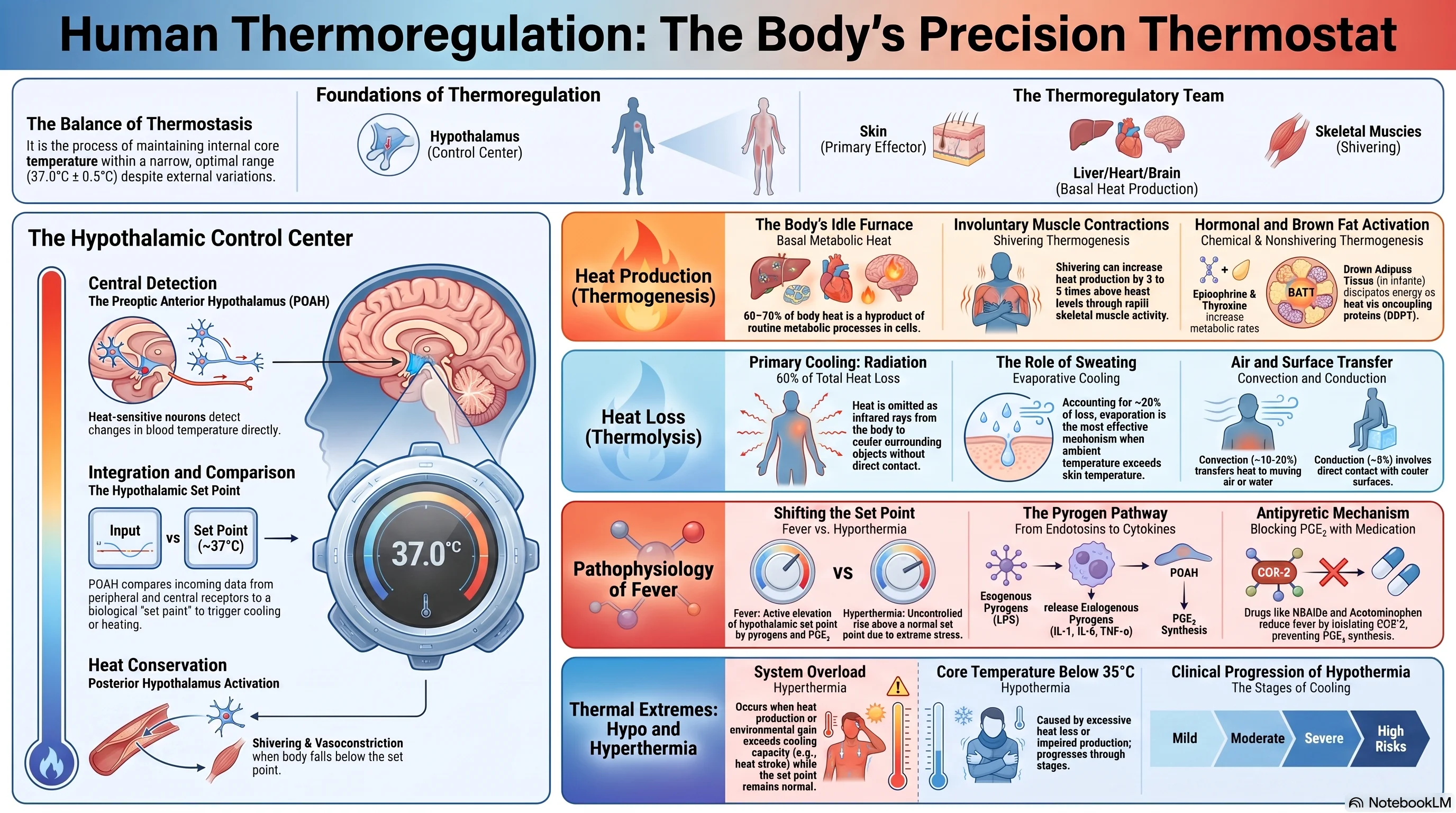
Thermoregulation is the homeostatic process that maintains core body temperature within a narrow, optimal range (~37 °C ± 0.5 °C) despite environmental fluctuations. It is essential for enzyme function, membrane fluidity, and overall metabolism.
A. Core vs. Shell Temperature
| Compartment | Location | Normal Range | Characteristics |
|---|---|---|---|
| Core | Deep tissues (brain, thorax, abdomen) | 36.5–37.5 °C | Tightly regulated, stable |
| Shell | Skin and subcutaneous tissues | 28–36 °C | Varies with environment, cooler than core |
Measurement sites: oral (36.3–37.1 °C), rectal (36.7–37.5 °C – most accurate), tympanic, axillary, temporal artery.
B. Normal Variations
| Factor | Effect | Mechanism |
|---|---|---|
| Circadian rhythm | Lowest ~4–6 AM, highest ~4–6 PM (0.5–1 °C variation) | Endogenous biological clock |
| Age | Infants: unstable; Elderly: lower baseline, reduced regulation | Immature/declining thermoregulation |
| Gender | Women ~0.3–0.5 °C higher; rises during luteal phase | Progesterone effect |
| Exercise | May rise to 39–40 °C | Muscle heat production |
| Meals | Slight increase | Thermic effect of food |
| Emotion | Slight increase | Sympathetic activation |
2. Mechanisms of Body Temperature Regulation
Body temperature is determined by the balance between heat production (thermogenesis) and heat loss (thermolysis), controlled by the hypothalamus.
A. Heat Production (Thermogenesis)
| Mechanism | Description | Regulation |
|---|---|---|
| Basal metabolism | Heat from routine cellular activity (liver, brain, heart) – ~60% of resting heat | Not regulated for temperature |
| Muscular activity | Voluntary exercise and involuntary shivering | Shivering: hypothalamus; Exercise: voluntary |
| Shivering thermogenesis | Rhythmic skeletal muscle contractions, increases heat 4–5× | Posterior hypothalamus → motor pathways |
| Non‑shivering thermogenesis | Uncoupling protein‑1 (UCP1) in brown adipose tissue (BAT) dissipates proton gradient as heat | Sympathetic β₃ stimulation; significant in infants, small amounts in adults |
| Chemical thermogenesis | Hormones (thyroxine, epinephrine, norepinephrine) increase metabolic rate | Thyroid: long‑term cold adaptation; Catecholamines: acute |
- Thyroid hormones (T₃/T₄) enhance Na⁺/K⁺‑ATPase activity and mitochondrial enzymes, raising basal metabolic rate over days to weeks.
- Brown adipose tissue (BAT): Located in neck, supraclavicular, paravertebral regions; activated by cold exposure via sympathetic nerves.
B. Heat Loss (Thermolysis)
| Route | Contribution at rest | Mechanism | Modulation |
|---|---|---|---|
| Radiation | ~60% | Emission of infrared rays to cooler surroundings | Depends on skin–environment gradient |
| Convection | ~15% | Transfer to moving air/water | Increased by wind; body can use piloerection (minimal in humans) |
| Conduction | ~3% | Direct transfer to cooler objects in contact | Enhanced by cold surfaces (water immersion) |
| Evaporation | ~22% | Heat loss when sweat vaporises | Highly regulated – each mL removes ~0.58 kcal |
Sweating:
- Eccrine sweat glands innervated by sympathetic cholinergic fibres.
- Secretion is hypotonic (water, Na⁺, Cl⁻, K⁺, lactate).
- Up to 1–2 L/hour during heavy exercise; inefficient in high humidity.
Cutaneous blood flow:
- Vasodilation (active): NO, bradykinin, VIP release → increased skin blood flow (up to 8 L/min) → heat loss.
- Vasoconstriction (sympathetic α‑adrenergic): reduces skin blood flow near zero in cold.
Countercurrent heat exchange: In limbs, arteries and veins run parallel; warm arterial blood pre‑warms cool venous blood, reducing heat loss from extremities.
C. Thermal Balance Equation
Heat storage = Heat production – Heat loss
- If production > loss → temperature rises.
- If production < loss → temperature falls.
- At equilibrium, production = loss.
3. The Hypothalamus: The Body’s Thermostat
The hypothalamus integrates thermal information and orchestrates efferent responses to maintain core temperature at the set point (~37 °C).
| Component | Structure | Function |
|---|---|---|
| Sensors | Peripheral thermoreceptors (skin) and central thermoreceptors (hypothalamus, spinal cord, abdominal organs) | Detect temperature changes |
| Integrator | Preoptic anterior hypothalamus (POAH) | Compares actual temperature to set point; coordinates responses |
| Effectors | Sweat glands, cutaneous vessels, skeletal muscle, endocrine glands | Execute heat loss or heat production/conservation |
Temperature sensors:
- Peripheral: Cold receptors (Aδ fibres, peak ~25 °C) and warm receptors (C fibres, peak ~43 °C) in skin → spinothalamic tract → thalamus → POAH.
- Central: Warm‑sensitive neurons in POAH increase firing with heat; cold‑sensitive neurons increase firing with cooling.
Set point mechanism:
- Determined by balance of warm‑ and cold‑sensitive neurons in POAH.
- If actual temperature < set point → heat conservation/production activated.
- If actual temperature > set point → heat loss activated.
Efferent pathways:
| Response | Pathway |
|---|---|
| Vasoconstriction | POAH → posterior hypothalamus → sympathetic (α‑adrenergic) → cutaneous vessels |
| Vasodilation | Inhibition of sympathetic tone; active vasodilation via NO, VIP |
| Sweating | POAH → anterior hypothalamus → sympathetic cholinergic fibres → sweat glands |
| Shivering | POAH → posterior hypothalamus → rubro‑/reticulospinal tracts → α‑motor neurons |
| Behavioral | Hypothalamus → cerebral cortex → conscious sensation → voluntary adjustments |
Responses to cold:
- Cutaneous vasoconstriction, piloerection (minimal in humans), shivering, non‑shivering thermogenesis (BAT), increased thyroid hormone (chronic), behavioral (seek warmth, add clothing).
Responses to heat:
- Cutaneous vasodilation, sweating, behavioural (seek shade, reduce activity), inhibition of heat production.
4. Pathophysiology of Fever
Fever is an elevation of body temperature resulting from a raised hypothalamic set point, usually due to infection or inflammation.
Distinction from hyperthermia:
| Feature | Fever | Hyperthermia |
|---|---|---|
| Set point | Increased | Normal |
| Mechanism | Pyrogens → PGE₂ → raised set point | Heat gain exceeds loss; impaired thermoregulation |
| Body temperature | Rises to new set point | Rises above set point |
| Response to antipyretics | Yes (lowers set point) | No |
| Examples | Infection, autoimmune disease | Heat stroke, malignant hyperthermia, drug toxicity |
A. Pyrogens
- Exogenous pyrogens: Microbial products (LPS from Gram‑negative bacteria, lipoteichoic acid, viral nucleic acids).
- Endogenous pyrogens (cytokines): IL‑1 (primary), IL‑6, TNF‑α, interferons – released by macrophages/monocytes.
B. Mechanism of Fever
- Exogenous pyrogen binds TLRs on immune cells.
- Release of endogenous pyrogens (IL‑1, IL‑6, TNF‑α).
- Cytokines act on circumventricular organs (OVLT) → induce COX‑2 in hypothalamic endothelium → PGE₂ synthesis.
- PGE₂ binds EP3 receptors on warm‑sensitive neurons in POAH → decreases their firing rate → set point rises.
- Body perceives actual temperature as “cold” and activates heat conservation (vasoconstriction) and production (shivering, rigors).
- Temperature rises to the new set point.
- When pyrogenic stimulus ends, PGE₂ falls, set point returns to normal; heat loss mechanisms (vasodilation, sweating) cause defervescence.
Stages of fever:
- Prodrome: malaise, aches.
- Chill (rigor): shivering, vasoconstriction, feeling cold.
- Febrile plateau: temperature stable at elevated set point, flushed skin.
- Defervescence: sweating, vasodilation, temperature falls.
C. Benefits and Risks of Fever
- Benefits: enhanced immune function (leukocyte migration, phagocytosis, interferon), inhibition of pathogen growth.
- Risks: dehydration, increased metabolic demand (12% per °C), febrile seizures (children), exacerbation of pre‑existing conditions.
D. Antipyretic Drugs
| Drug Class | Mechanism | Examples |
|---|---|---|
| NSAIDs | Inhibit COX‑2 (and COX‑1) → ↓ PGE₂ synthesis | Ibuprofen, aspirin, naproxen |
| Acetaminophen (paracetamol) | Weak COX inhibitor; central action, possibly COX‑3 | Paracetamol |
| Corticosteroids | Inhibit cytokine production (rarely used for fever alone) | Prednisone |
Physical cooling (sponging, ice packs) is not recommended for fever because it induces shivering and discomfort, working against the raised set point. It is reserved for hyperthermia or extreme hyperpyrexia.
5. Pathophysiology of Hypothermia
Hypothermia is a core temperature <35 °C (95 °F) due to excessive heat loss, insufficient heat production, or impaired thermoregulation.
A. Causes
| Category | Examples |
|---|---|
| Environmental | Cold water immersion, inadequate clothing |
| Metabolic/endocrine | Hypothyroidism, hypoglycaemia, adrenal insufficiency |
| Drug‑induced | Alcohol (vasodilation, impaired judgement), sedatives, opioids, antipsychotics |
| Neurological | Stroke, spinal cord injury, hypothalamic lesions |
| Sepsis | Impaired thermoregulation (especially in elderly) |
| Age extremes | Infants (large surface area/mass ratio, limited shivering); Elderly (decreased muscle mass, medications, impaired vasoconstriction) |
B. Stages of Hypothermia
| Stage | Core Temperature | Clinical Features |
|---|---|---|
| Mild | 32–35 °C (90–95 °F) | Shivering, tachycardia, tachypnoea, cold diuresis, confusion, ataxia |
| Moderate | 28–32 °C (82–90 °F) | Shivering ceases (<32 °C), bradycardia, bradypnoea, hypotension, dilated pupils, stupor, Osborn J wave on ECG |
| Severe | <28 °C (<82 °F) | Coma, areflexia, apnoea, ventricular fibrillation/asystole, may appear dead |
| Profound | <20 °C (<68 °F) | No vital signs; paradoxical undressing; highest risk of cardiac arrest |
Cold diuresis: vasoconstriction increases central blood volume → suppression of ADH → diuresis → potential hypovolaemia.
Osborn (J) wave: positive deflection at J point (QRS‑ST junction), characteristic of moderate‑severe hypothermia.
C. Management
- Remove from cold environment, remove wet clothing.
- Mild: passive external rewarming (blankets, warm environment).
- Moderate: active external rewarming (forced‑air warming, warm water bottles).
- Severe: active internal rewarming (warmed IV fluids 40–42 °C, warm humidified O₂, peritoneal/pleural lavage, extracorporeal rewarming).
- CPR may be required; the adage “a patient is not dead until warm and dead” applies. Complications: afterdrop (core cooling during rewarming), rewarming shock (vasodilation → hypotension), arrhythmias.
6. Clinical and Pharmaceutical Relevance
A. Drugs Affecting Thermoregulation
| Drug Class | Effect | Mechanism |
|---|---|---|
| Antipyretics (NSAIDs, paracetamol) | ↓ Fever | Inhibit COX → ↓ PGE₂ → lower set point |
| Antipsychotics (phenothiazines) | Hypothermia or hyperthermia | Block dopamine; impair hypothalamic regulation; α‑blockade → vasodilation |
| Anaesthetics (volatile) | Hypothermia | Impair thermoregulation; vasodilation; decrease metabolism |
| Succinylcholine + volatile anaesthetics | Malignant hyperthermia | Trigger uncontrolled Ca²⁺ release from SR via abnormal RyR1 |
| Sympathomimetics (cocaine, MDMA, amphetamines) | Hyperthermia | Increased activity; vasoconstriction; impaired heat dissipation |
| Anticholinergics (atropine, tricyclics) | Hyperthermia | Inhibit sweating |
| Alcohol (acute) | Hypothermia | Vasodilation; impaired judgement; decreased shivering |
| Beta‑blockers | May impair cold‑induced vasodilation | Block β‑receptors |
B. Malignant Hyperthermia (MH)
- Trigger: volatile anaesthetics (halothane, sevoflurane) or succinylcholine in genetically susceptible individuals (RYR1, CACNA1S mutations).
- Pathophysiology: uncontrolled Ca²⁺ release from SR via abnormal ryanodine receptor → sustained muscle contraction → massive heat production, rhabdomyolysis, hyperkalaemia, acidosis.
- Clinical: masseter rigidity, tachycardia, rapid temperature rise (up to 1 °C/5 min), metabolic acidosis, hyperkalaemia, elevated CK.
- Treatment: dantrolene (inhibits RyR, reduces Ca²⁺ release), stop triggering agent, hyperventilate with 100% O₂, active cooling, treat complications.
C. Heat Stroke
- Definition: core temperature >40 °C with neurological dysfunction, due to overwhelmed thermoregulation.
- Types:
- Classic (non‑exertional): elderly, chronic illness, medications; gradual onset, anhidrotic skin.
- Exertional: young individuals during strenuous exercise; rapid onset, sweating may persist.
- Pathophysiology: direct cellular toxicity, SIRS, endothelial injury, DIC, multi‑organ failure.
- Clinical: hyperthermia, CNS dysfunction (confusion, seizures, coma), cardiovascular collapse, hepatic necrosis, acute kidney injury.
- Treatment: rapid cooling (ice water immersion, evaporative cooling, cold IV fluids), supportive care.
Tables
Table 1: Heat Production vs. Heat Loss Mechanisms
| Heat Production | Heat Loss |
|---|---|
| Basal metabolism (60%) | Radiation (~60%) |
| Shivering (4–5× increase) | Convection (~15%) |
| Non‑shivering thermogenesis (BAT, UCP1) | Conduction (~3%) |
| Chemical thermogenesis (thyroid, catecholamines) | Evaporation (~22% – sweating) |
| Voluntary muscle activity | Respiratory evaporation (insensible) |
Table 2: Thermoregulatory Responses to Heat and Cold
| Condition | Effector | Response | Mechanism |
|---|---|---|---|
| Cold | Cutaneous vessels | Vasoconstriction | Sympathetic α‑adrenergic |
| Skeletal muscle | Shivering | Posterior hypothalamus → motor pathways | |
| BAT | Non‑shivering thermogenesis | Sympathetic β₃ → UCP1 | |
| Thyroid | ↑ T₃/T₄ (chronic) | TRH → TSH | |
| Behaviour | Seek warmth, add clothing | Conscious | |
| Heat | Cutaneous vessels | Vasodilation | Inhibited sympathetic tone; active vasodilation (NO) |
| Sweat glands | Sweating | Sympathetic cholinergic | |
| Behaviour | Seek shade, reduce activity | Conscious | |
| Metabolism | Decreased heat production | Reduced muscle tone |
Table 3: Fever vs. Hyperthermia
| Feature | Fever | Hyperthermia |
|---|---|---|
| Set point | Increased | Normal |
| Mechanism | Pyrogens → PGE₂ → raised set point | Heat gain > heat loss; impaired thermoregulation |
| Response to antipyretics | Yes | No |
| Response to cooling | May cause shivering (defends elevated set point) | Effective (no defence) |
| Examples | Infection, inflammation | Heat stroke, malignant hyperthermia, drug toxicity |
Table 4: Stages of Hypothermia
| Stage | Temperature | Shivering | Consciousness | Cardiovascular | Management |
|---|---|---|---|---|---|
| Mild | 32–35 °C | Present | Confused | Tachycardia | Passive external rewarming |
| Moderate | 28–32 °C | Absent | Stuporous | Bradycardia, J wave | Active external rewarming |
| Severe | <28 °C | Absent | Coma | Hypotension, VF/asystole | Active internal rewarming, CPR |
Exam Angle
Short Answer Questions
- Differentiate between fever and hyperthermia.
- How does the hypothalamus regulate body temperature?
- What are endogenous pyrogens? Give examples.
- List the mechanisms of heat loss from the body.
- Define hypothermia and list its stages.
Essay Questions
- Explain the pathophysiology of fever, including the role of pyrogens, prostaglandins, and the hypothalamic set point.
- Describe the mechanisms of body temperature regulation, including heat production and heat loss.
- Discuss the pathophysiology and clinical management of hypothermia.
Viva / Short Notes
- Role of the hypothalamus as thermostat.
- Endogenous pyrogens (IL‑1, IL‑6, TNF‑α).
- Shivering thermogenesis.
- Cutaneous vasodilation and vasoconstriction.
- Mechanism of action of antipyretic drugs.
- Malignant hyperthermia.
- Heat stroke.
Summary Box
- Body temperature is maintained by a balance between heat production (basal metabolism, shivering, non‑shivering thermogenesis, chemical thermogenesis) and heat loss (radiation, convection, conduction, evaporation).
- The hypothalamus (POAH) acts as the thermostat, comparing actual temperature to a set point (~37 °C) and initiating appropriate responses via autonomic, somatic, and endocrine pathways.
- Fever results from an increased set point driven by pyrogens (exogenous → endogenous cytokines → PGE₂ synthesis via COX‑2). Antipyretics (NSAIDs, paracetamol) lower the set point by inhibiting COX.
- Hyperthermia differs from fever: the set point is normal, but thermoregulation is overwhelmed (e.g., heat stroke, malignant hyperthermia).
- Hypothermia is a core temperature <35 °C, staged from mild to severe, and is managed by passive/active rewarming. Severe cases require internal rewarming and prolonged CPR.
- Pharmacy relevance: many drugs affect thermoregulation (antipyretics, antipsychotics, anaesthetics, sympathomimetics, anticholinergics). Malignant hyperthermia is a life‑threatening pharmacogenetic emergency treated with dantrolene.
References
- Hall, J. E. (2021). Guyton and Hall Textbook of Medical Physiology (14th ed.). Elsevier. (Chapter 74: Body Temperature Regulation and Fever)
- Barrett, K. E., et al. (2019). Ganong’s Review of Medical Physiology (26th ed.). McGraw‑Hill.
- Costanzo, L. S. (2024). BRS Physiology (7th ed.). Wolters Kluwer.
- Widmaier, E. P., et al. (2023). Vander’s Human Physiology (16th ed.). McGraw‑Hill.
- Bouchama, A., & Knochel, J. P. (2002). Heat stroke. N Engl J Med, 346(25), 1978‑1988.
- Brown, D. J., & Brugger, H. (2012). Accidental hypothermia. N Engl J Med, 367(20), 1930‑1938.
- Rosenberg, H., et al. (2015). Malignant hyperthermia: a review. Orphanet J Rare Dis, 10, 93.
End of unit 7 of 7
Check what you've learned
5 quick questions from this unit. They're optional: try them if you like, then mark the unit as read and move on.