

Unit 5: The Blood Cells
RBCs, WBCs, platelets, haemostasis & blood groups.
Prepared by the PharmaWallah student team. Verify results with your textbook or instructor. Report an error · Editorial policy
Unit 5: The Blood Cells
Learning Objectives
- Describe the structure, function, and life cycle of red blood cells, including erythropoiesis and hemoglobin metabolism.
- Identify the types of white blood cells, their genesis, and their specific roles in immunity.
- Explain the functions of T‑cells, B‑cells, memory cells, and antibodies in the immune response.
- Describe the formation of platelets, the platelet plug, and the mechanisms of blood coagulation (intrinsic and extrinsic pathways).
- Explain blood typing, agglutinogens, Rh blood types, and the composition of plasma and serum.
- Understand the clinical significance of anemia, leukopenia, and pus formation.
Core Content
1. Red Blood Cells (Erythrocytes)
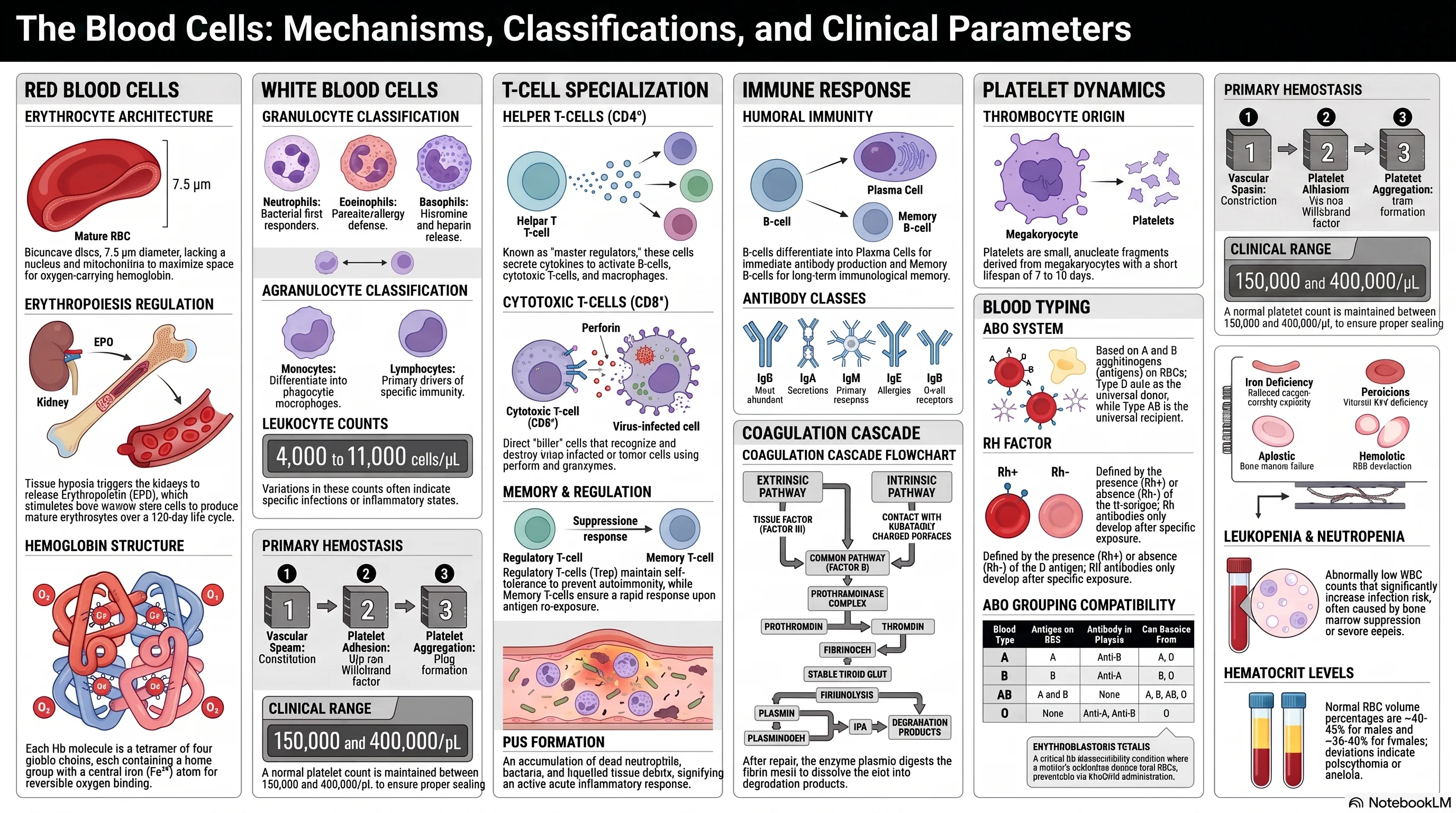
Red blood cells (RBCs) are the most abundant blood cells, specialised for oxygen and carbon dioxide transport.
A. Structure and Characteristics
| Feature | Description | Functional Significance |
|---|---|---|
| Shape | Biconcave disc | Maximises surface area for gas exchange; allows deformation through narrow capillaries |
| Size | ~7.5 µm diameter | Slightly larger than capillary diameter, requiring deformation |
| Nucleus | Absent (extruded during maturation) | Maximises space for haemoglobin; prevents cell division |
| Organelles | Absent (no mitochondria, ribosomes, ER) | Glycolysis only – no consumption of transported O₂ |
| Haemoglobin | ~280 million molecules per cell | Binds and transports O₂ and CO₂ |
| Lifespan | ~120 days | |
| Count | Males: 4.5–6.0 million/µL; Females: 4.0–5.5 million/µL |
Membrane skeleton: Spectrin, actin, ankyrin – defects cause hereditary spherocytosis.
B. Erythropoiesis (Formation of RBCs)
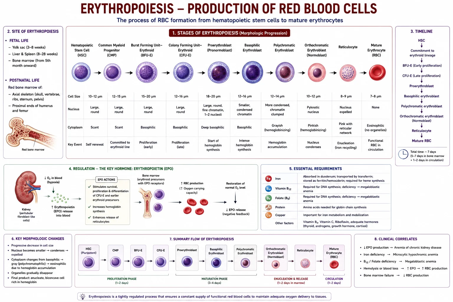
Erythropoiesis occurs in red bone marrow.
Sites throughout life:
- Fetus: Yolk sac → liver & spleen → bone marrow
- Adult: Flat bones (sternum, ribs, pelvis, vertebrae, skull) and proximal ends of long bones.
Stages of erythropoiesis:
- Pluripotent haematopoietic stem cell → 2. Myeloid stem cell → 3. Proerythroblast (large, nucleated) → 4. Basophilic erythroblast (haemoglobin synthesis begins) → 5. Polychromatophilic erythroblast (haemoglobin increases) → 6. Orthochromatic erythroblast (nucleus condenses and is extruded) → 7. Reticulocyte (released into blood; residual RNA, matures in 1–2 days) → 8. Mature erythrocyte
Regulation by Erythropoietin (EPO):
- Produced mainly by kidneys (peritubular fibroblasts) in response to hypoxia.
- Hypoxia‑inducible factor (HIF‑1α) stimulates EPO gene transcription.
- EPO acts on bone marrow CFU‑E and BFU‑E, promoting proliferation, differentiation, and inhibiting apoptosis.
- Negative feedback: increased RBC mass → improved O₂ delivery → decreased EPO.
Factors required for erythropoiesis:
| Factor | Role | Deficiency result |
|---|---|---|
| Iron | Heme synthesis | Microcytic hypochromic anaemia |
| Vitamin B₁₂ | DNA synthesis | Megaloblastic anaemia |
| Folate | DNA synthesis | Megaloblastic anaemia |
| Vitamin B₆ | Heme synthesis (ALA synthase cofactor) | Sideroblastic anaemia |
| Copper | Iron mobilisation | Microcytic anaemia |
C. Haemoglobin (Hb)
- Tetramer of four globin chains: 2α + 2β (HbA), each with a heme group (protoporphyrin IX + Fe²⁺).
- Each heme binds one O₂; cooperative binding (sigmoidal O₂ dissociation curve).
- Types: HbA (α₂β₂, >95%), HbA₂ (α₂δ₂, 2–3.5%), HbF (α₂γ₂, fetal – higher O₂ affinity).
Normal values: Males 13.5–17.5 g/dL; Females 12.0–16.0 g/dL.
Haemoglobin breakdown (RBC lifecycle):
- Senescent RBCs phagocytosed by macrophages in spleen/liver/bone marrow.
- Globin hydrolysed to amino acids → recycled.
- Heme oxygenase cleaves heme → biliverdin, CO, Fe²⁺.
- Biliverdin → bilirubin (unconjugated, bound to albumin in blood).
- Liver conjugates bilirubin → excreted in bile.
- Gut bacteria convert to urobilinogen → partly excreted in faeces (stercobilin) and urine (urobilin). Jaundice: Pre‑hepatic (haemolytic, unconjugated), Hepatic (impaired conjugation), Post‑hepatic (obstructive, conjugated).
2. White Blood Cells (Leukocytes)
White blood cells are the immune system’s mobile defence units. Normal count: 4,000–11,000/µL.
A. Classification
| Type | % of WBCs | Granules | Nucleus | Key Function |
|---|---|---|---|---|
| Granulocytes | ||||
| Neutrophils | 50–70% | Fine, pale | 3–5 lobes | Phagocytosis of bacteria; first responders; pus formation |
| Eosinophils | 1–4% | Coarse, red‑orange | 2 lobes | Parasite defence; allergic reactions |
| Basophils | <1% | Coarse, deep blue‑purple | 2 lobes | Release histamine, heparin; allergy & inflammation |
| Agranulocytes | ||||
| Lymphocytes | 20–40% | None (clear cytoplasm) | Round, dense | Specific immunity (T‑cells, B‑cells, NK cells) |
| Monocytes | 2–8% | None (fine azurophilic) | Kidney‑shaped | Phagocytosis; differentiate into macrophages and dendritic cells |
B. Leukopoiesis
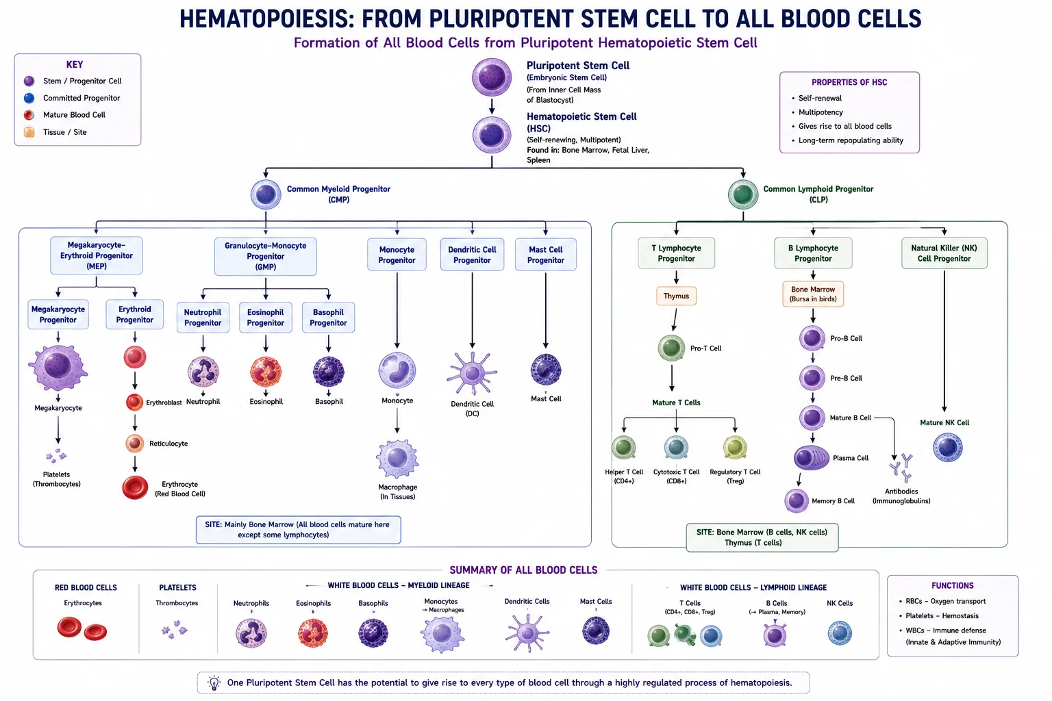
All blood cells arise from haematopoietic stem cells:
- Myeloid stem cell → granulocytes, monocytes, megakaryocytes, erythrocytes.
- Lymphoid stem cell → T‑lymphocytes, B‑lymphocytes, NK cells. Regulated by colony‑stimulating factors (G‑CSF, GM‑CSF, M‑CSF) and interleukins (IL‑3, IL‑5, IL‑7).
C. Roles of Different WBCs
- Neutrophils: Phagocytose bacteria; use oxidative burst (superoxide, H₂O₂) and enzymes. Dead neutrophils + bacteria + debris = pus.
- Eosinophils: Attack parasites (release major basic protein); modulate allergy.
- Basophils/Mast cells: Bind IgE; degranulate histamine, heparin, leukotrienes → vasodilation, bronchoconstriction.
- Monocytes/Macrophages: Phagocytosis, antigen presentation, cytokine secretion (IL‑1, TNF‑α). Named in tissues: Kupffer cells (liver), microglia (brain), osteoclasts (bone).
- Lymphocytes: Mediate adaptive immunity.
3. T‑Cells and B‑Cells in Immunity
A. T‑Cells (Cell‑mediated immunity)
Develop in thymus; recognise antigen presented by MHC molecules.
| T‑Cell Subset | Surface Marker | Function |
|---|---|---|
| Helper T‑cells (Th) | CD4+ (MHC‑II restricted) | Orchestrate immune responses by secreting cytokines: Th1 (activate macrophages), Th2 (help B‑cells, allergy), Th17 (extracellular bacteria), Tfh (germinal centre B‑cell help), Treg (suppress immunity) |
| Cytotoxic T‑cells (Tc) | CD8+ (MHC‑I restricted) | Kill virus‑infected/tumour cells via perforin, granzymes, Fas ligand |
| Regulatory T‑cells (Treg) | CD4+ CD25+ FoxP3+ | Maintain self‑tolerance; prevent autoimmunity |
| Memory T‑cells | CD4+ or CD8+ | Long‑lived; rapid response upon re‑exposure |
Activation: Requires antigen presentation + co‑stimulation (CD28–CD80/86). Results in clonal expansion and differentiation.
B. B‑Cells and Antibodies (Humoral immunity)
Mature in bone marrow; each B‑cell expresses a unique surface immunoglobulin (BCR).
- Upon activation (with T‑cell help), differentiate into:
- Plasma cells: antibody factories (abundant rough ER).
- Memory B‑cells: long‑lived, rapid secondary response.
Antibody (Immunoglobulin) Classes:
| Class | Structure | Serum % | Key Functions |
|---|---|---|---|
| IgG | Monomer | 75% | Main antibody in secondary response; opsonisation; complement; crosses placenta |
| IgA | Monomer (serum) / Dimer (secretory) | 15% | Mucosal immunity (saliva, tears, breast milk) |
| IgM | Pentamer | 10% | Primary response; most efficient complement activator; BCR (monomer) |
| IgE | Monomer | <0.1% | Binds mast cells/basophils; allergy; anti‑parasite |
| IgD | Monomer | <1% | BCR (with IgM); B‑cell activation |
Antibody structure: Y‑shaped, 2 heavy + 2 light chains; variable region binds antigen; constant region mediates effector functions.
C. Memory Cells
- Central memory T‑cells (Tcm): in lymphoid organs; proliferate upon re‑stimulation.
- Effector memory T‑cells (Tem): in tissues; immediate effector function.
- Memory B‑cells: express high‑affinity, class‑switched BCR; rapidly become plasma cells.
- Immunological memory is the basis of vaccination.
4. Pus Formation
Pus is a thick, yellowish fluid consisting of:
- Dead and dying neutrophils
- Necrotic tissue debris
- Living and dead bacteria
- Inflammatory exudate
Formation: Neutrophils are attracted by chemotactic signals → phagocytose pathogens → die (apoptosis/necrosis) → release proteolytic enzymes → liquefaction of tissue. Accumulated mass may form an abscess if walled off.
5. Anaemia and Leukopenia
A. Anaemia (reduced O₂‑carrying capacity)
Classified by RBC size (MCV) and mechanism:
| Type | Causes | Lab Features |
|---|---|---|
| Microcytic hypochromic (MCV <80 fL) | Iron deficiency (chronic blood loss, poor diet); Thalassaemia; Anaemia of chronic disease; Sideroblastic anaemia | Low ferritin (iron deficiency); abnormal Hb electrophoresis (thalassaemia) |
| Macrocytic (MCV >100 fL) | Megaloblastic (B₁₂/folate deficiency); Non‑megaloblastic (liver disease, alcohol, hypothyroidism) | Hypersegmented neutrophils (megaloblastic); low B₁₂/folate |
| Normocytic normochromic (MCV 80–100 fL) | Acute blood loss; Haemolytic anaemia; Bone marrow failure; Anaemia of chronic disease (early) | Increased reticulocytes (haemolysis); pancytopenia (marrow failure) |
B. Leukopenia (WBC count <4,000/µL)
- Neutropenia: chemotherapy, radiation, bone marrow disorders, severe infection, drugs (clozapine). Increases risk of bacterial/fungal infections.
- Lymphopenia: HIV/AIDS, immunosuppressive drugs, corticosteroids. Risk of opportunistic infections.
- Pancytopenia: aplastic anaemia, myelodysplasia, hypersplenism.
6. Platelets (Thrombocytes)
Platelets are anucleate cell fragments from megakaryocytes. Normal count: 150,000–450,000/µL; lifespan 7–10 days.
A. Formation (Thrombopoiesis)
- Regulated by thrombopoietin (TPO) from liver; TPO receptors (c‑Mpl) on megakaryocytes and platelets.
- Megakaryocytes undergo endomitosis → polyploid → extend proplatelets into sinusoids → shed platelets.
Platelet structure:
- Glycocalyx with glycoproteins (GP Ib‑IX‑V, GP IIb/IIIa) for adhesion/aggregation.
- α‑granules: fibrinogen, vWF, PDGF, PF4.
- Dense granules: ADP, ATP, serotonin, Ca²⁺.
- Open canalicular system for granule release.
B. Platelet Plug Formation (Primary Haemostasis)
- Vascular spasm: vasoconstriction (endothelin, serotonin).
- Adhesion: Platelet GP Ib binds vWF on exposed collagen.
- Activation: Shape change, release of ADP, thromboxane A₂ (TXA₂), serotonin.
- Aggregation: Fibrinogen bridges activated GP IIb/IIIa receptors → platelet plug. Key drugs: Aspirin inhibits COX‑1 → ↓ TXA₂; Clopidogrel blocks P2Y₁₂ (ADP receptor).
7. Blood Coagulation (Secondary Haemostasis)
Conversion of soluble fibrinogen → insoluble fibrin via a cascade of serine protease zymogens. Most factors are synthesised in the liver; Vitamin K is required for factors II, VII, IX, X.
A. Coagulation Factors (Roman numerals)
| Factor | Name | Pathway | Vitamin K‑dependent |
|---|---|---|---|
| I | Fibrinogen | Common | No |
| II | Prothrombin | Common | Yes |
| III | Tissue factor | Extrinsic | No |
| IV | Calcium | All | No |
| V | Proaccelerin | Common | No |
| VII | Proconvertin | Extrinsic | Yes |
| VIII | Anti‑haemophilic factor | Intrinsic | No |
| IX | Christmas factor | Intrinsic | Yes |
| X | Stuart‑Prower factor | Common | Yes |
| XI | Plasma thromboplastin antecedent | Intrinsic | No |
| XII | Hageman factor | Intrinsic | No |
| XIII | Fibrin‑stabilising factor | Common | No |
B. Pathways
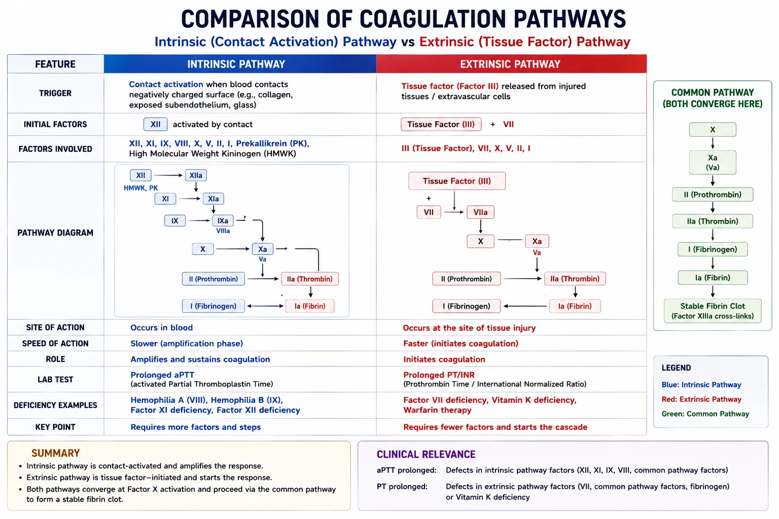
Extrinsic pathway (initiated by tissue factor from damaged cells):
TF + VIIa → activates X → common pathway. Fast (seconds).
Intrinsic pathway (contact activation, e.g., exposed collagen):
XII → XIIa → XIa → IXa (+ VIIIa) → activates X. Slower (minutes).
Common pathway:
Xa + Va + Ca²⁺ + phospholipid (prothrombinase complex) → Prothrombin (II) → Thrombin (IIa).
Thrombin converts fibrinogen (I) to fibrin monomers; activates XIII → cross‑links fibrin → stable clot.
Thrombin also: Activates platelets; enhances factors V, VIII, XI (positive feedback); activates protein C (negative feedback).
C. Regulation & Fibrinolysis
- Natural anticoagulants: Antithrombin III (enhanced by heparin), Protein C + S (inactivate Va, VIIIa), TFPI (inhibits VIIa/TF).
- Fibrinolysis: Tissue plasminogen activator (tPA) converts plasminogen (in clot) to plasmin → digests fibrin → fibrin degradation products (e.g., D‑dimer). α₂‑antiplasmin and PAI‑1 limit the process.
- Clinical: PT/INR monitors warfarin; aPTT monitors heparin; D‑dimer detects thrombosis.
8. Blood Types
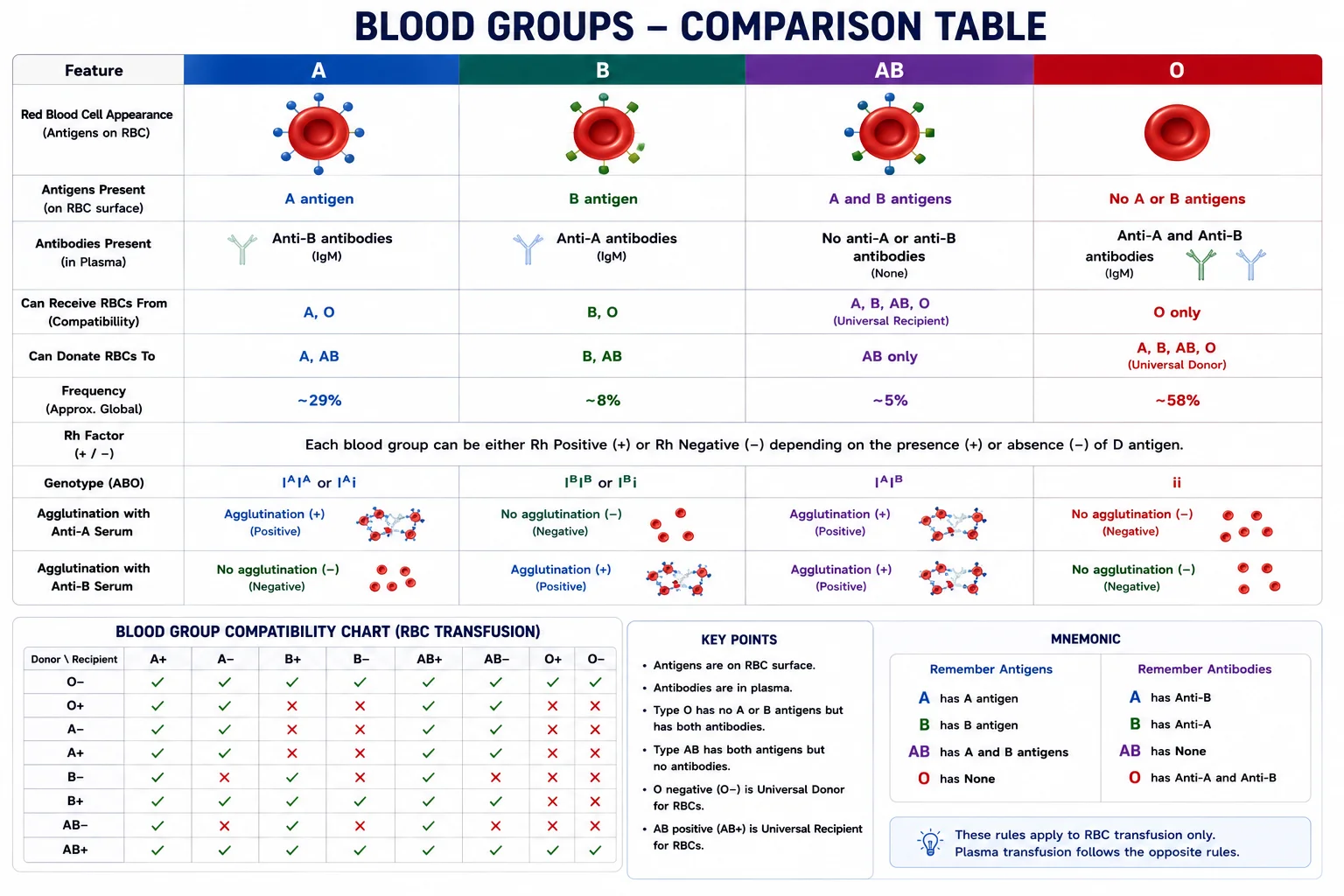
A. ABO System
| Blood Group | RBC Antigens | Plasma Antibodies | Can Donate To | Can Receive From |
|---|---|---|---|---|
| A | A | Anti‑B | A, AB | A, O |
| B | B | Anti‑A | B, AB | B, O |
| AB | A & B | None (universal recipient) | AB | A, B, AB, O |
| O | None (H antigen) | Anti‑A, Anti‑B (universal donor) | A, B, AB, O | O |
B. Rh System
- Rh‑positive: D antigen present (85%); Rh‑negative: absent (15%).
- Anti‑D antibodies are not naturally occurring; produced after exposure (transfusion or pregnancy).
- Erythroblastosis fetalis: Rh‑negative mother with Rh‑positive fetus → maternal anti‑D IgG crosses placenta in subsequent pregnancies → haemolytic disease of newborn.
- Prevention: Rh immunoglobulin (RhoGAM) at 28 weeks and after delivery.
9. Plasma and Serum
| Feature | Plasma | Serum |
|---|---|---|
| Definition | Liquid portion of unclotted blood (anticoagulant added) | Liquid after blood has clotted (no anticoagulant) |
| Fibrinogen | Present | Absent (consumed as fibrin) |
| Clotting factors | All present | Absent (consumed) |
| Use | Coagulation studies, fresh frozen plasma | Chemistry tests (electrolytes, proteins, etc.) |
| Colour | Straw‑coloured | Slightly lighter |
Plasma proteins:
- Albumin (55–60%): oncotic pressure, transport.
- Globulins (35–40%): α, β (transport), γ (immunoglobulins).
- Fibrinogen (4%): coagulation.
Tables
Table 1: Summary of Clotting Pathways
| Pathway | Trigger | Key Activated Factors | Speed |
|---|---|---|---|
| Extrinsic | Tissue factor (TF) | VII, X | Seconds |
| Intrinsic | Contact with collagen | XII, XI, IX, VIII, X | Minutes |
| Common | Factor Xa from either pathway | II (prothrombin), I (fibrinogen), XIII | – |
Table 2: Normal Haematological Values
| Parameter | Normal Range |
|---|---|
| RBC count (male) | 4.5–6.0 million/µL |
| Haemoglobin (male) | 13.5–17.5 g/dL |
| Haematocrit (male) | 40–54% |
| WBC count | 4,000–11,000/µL |
| Platelet count | 150,000–450,000/µL |
| Reticulocytes | 0.5–1.5% of RBCs |
Exam Angle
Short Answer Questions
- Describe the regulation of erythropoiesis by erythropoietin.
- Differentiate between the extrinsic and intrinsic coagulation pathways.
- Compare the functions of neutrophils, eosinophils, and basophils.
- What is the difference between plasma and serum?
- Explain the role of memory cells in immunity.
Essay Questions
- Discuss the formation, structure, and breakdown of haemoglobin, including the fate of its components.
- Describe the mechanisms of haemostasis, including platelet plug formation and the coagulation cascade.
- Explain the ABO and Rh blood group systems and their clinical significance in transfusion and pregnancy.
Viva / Short Notes
- Erythropoiesis stages.
- Bilirubin metabolism and jaundice.
- Platelet adhesion and aggregation.
- Fibrinolysis.
- Functions of different immunoglobulin classes.
Summary Box
- Red blood cells are biconcave, anucleate, filled with haemoglobin (120‑day lifespan). Erythropoiesis is driven by EPO from kidneys in response to hypoxia. Old RBCs are broken down: heme → bilirubin; iron recycled.
- White blood cells protect against infection. Granulocytes (neutrophils, eosinophils, basophils) act non‑specifically; lymphocytes (T‑cells, B‑cells, NK cells) mediate specific immunity. Pus is an accumulation of dead neutrophils and debris.
- T‑cells mediate cellular immunity (helper CD4+, cytotoxic CD8+, regulatory). B‑cells produce antibodies (IgG, IgA, IgM, IgE, IgD). Memory cells enable rapid secondary responses.
- Platelets form the primary haemostatic plug (adhesion → activation → aggregation). Secondary haemostasis involves the coagulation cascade (extrinsic, intrinsic, common pathways) culminating in fibrin clot stabilisation. Clot is removed by fibrinolysis (plasmin).
- Blood groups are determined by ABO antigens (naturally occurring antibodies) and Rh factor. Rh incompatibility can cause haemolytic disease of newborn, prevented by RhoGAM.
- Plasma contains clotting factors (fibrinogen); serum is plasma without fibrinogen. Anaemia is reduced O₂‑carrying capacity; leukopenia increases infection risk.
References
- Hall, J. E. (2021). Guyton and Hall Textbook of Medical Physiology (14th ed.). Elsevier.
- Barrett, K. E., et al. (2019). Ganong’s Review of Medical Physiology (26th ed.). McGraw‑Hill.
- Costanzo, L. S. (2024). BRS Physiology (7th ed.). Wolters Kluwer.
- Hoffbrand, A. V. & Moss, P. A. H. (2020). Hoffbrand’s Essential Haematology (8th ed.). Wiley‑Blackwell.
- Katzung, B. G. (2018). Basic and Clinical Pharmacology (14th ed.). McGraw‑Hill.
End of unit 5 of 7
Check what you've learned
5 quick questions from this unit. They're optional: try them if you like, then mark the unit as read and move on.